模块 1. 化学品 1.1 产品标识符 :
睾酮 产品名称
1.2 鉴别的其他方法 trans-TestOSterone
17β-Hydroxy-3-oxo-4-andrOStene
17β-Hydroxy-4-andrOSten-3-one
4-AndrOSten-17β-ol-3-one
1.3 有关的确定了的物质或混合物的用途和建议不适合的用途 仅用于研发。不作为药品、家庭或其它用途。
模块 2. 危险性概述 2.1 GHS-分类 致癌性 (类别 1B)
生殖毒性 (类别 2)
2.2 GHS 标记要素,包括预防性的陈述 象形图
警示词 危险
危险申明
H350 可能致癌。
H361 怀疑对生育能力或胎儿造成伤害。
警告申明
预防措施
P201 在使用前获取特别指示。
P202 在读懂所有安全防范措施之前切勿操作。
P281 使用所需的个人防护设备。
事故响应
P308 + P313 如接触到或有疑虑:求医/ 就诊。
安全储存
P405 存放处须加锁。
废弃处置
P501 将内容物/ 容器处理到得到批准的废物处理厂。
只限于专业使用者。
2.3 其它危害物 - 无
模块 3. 成分/组成信息 3.1 物 质 : trans-TestOSterone
别名
17β-Hydroxy-3-oxo-4-andrOStene
17β-Hydroxy-4-andrOSten-3-one
4-AndrOSten-17β-ol-3-one
:
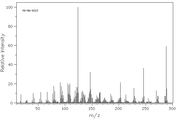
C19H28O2 分子式
: 288.42 g/mol
分子量
组分 浓度或浓度范围
TestOSterone
<=100%
化学文摘登记号(CAS
58-22-0 No.) 200-370-5
EC-编号
模块 4. 急救措施 4.1 必要的急救措施描述 一般的建议
请教医生。 向到现场的医生出示此安全技术说明书。
吸入
如果吸入,请将患者移到新鲜空气处。 如呼吸停止,进行人工呼吸。 请教医生。
皮肤接触
用肥皂和大量的
水冲洗。 请教医生。
眼睛接触
用
水冲洗眼睛作为预防措施。
食入
切勿给失去知觉者通过口喂任何东西。 用
水漱口。 请教医生。
4.2 主要症状和影响,急性和迟发效应 4.3 及时的医疗处理和所需的特殊处理的说明和指示 无数据资料
模块 5. 消防措施 5.1 灭火介质 灭火方法及灭火剂
用
水雾,抗
乙醇泡沫,干粉或
二氧化碳灭火。
5.2 源于此物质或混合物的特别的危害 碳氧化物
5.3 给消防员的建议 如必要的话,戴自给式呼吸器去救火。
5.4 进一步信息 无数据资料
模块 6. 泄露应急处理 6.1 作业人员防护措施、防护装备和应急处置程序 使用个人防护用品。 避免粉尘生成。 避免吸入蒸气、烟雾或气体。 保证充分的通风。
人员疏散到安全区域。 避免吸入粉尘。
6.2 环境保护措施 如能确保安全,可采取措施防止进一步的泄漏或溢出。 不要让产品进入下
水道。
6.3 泄漏化学品的收容、清除方法及所使用的处置材料 收集和处置时不要产生粉尘。 扫掉和铲掉。 放入合适的封闭的容器中待处理。
6.4 参考其他部分 丢弃处理请参阅第13节。
模块 7. 操作处置与储存 7.1 安全操作的注意事项 避免接触皮肤和眼睛。 避免形成粉尘和气溶胶。避免曝露:使用前需要获得专门的指导。
在有粉尘生成的地方,提供合适的排风设备。一般性的防火保护措施。
7.2 安全储存的条件,包括任何不兼容性 贮存在阴凉处。 使容器保持密闭,储存在干燥通风处。
7.3 特定用途 无数据资料
模块 8. 接触控制和个体防护 8.1 容许浓度 最高容许浓度
没有已知的国家规定的暴露极限。
8.2 暴露控制 适当的技术控制
根据良好的工业卫生和安全规范进行操作。 休息前和工作结束时洗手。
个体防护设备
眼/面保护
带有防护边罩的安全眼镜符合 EN166要求请使用经官方标准如NIOSH (美国) 或 EN 166(欧盟)
检测与批准的设备防护眼部。
皮肤保护
戴手套取 手套在使用前必须受检查。
请使用合适的方法脱除手套(不要接触手套外部表面),避免任何皮肤部位接触此产品.
使用后请将被污染过的手套根据相关法律法规和有效的实验室规章程序谨慎处理. 请清洗并吹干双手
所选择的保护手套必须符合EU的89/686/E
EC规定和从它衍生出来的EN 376标准。
完全接触
物料:
丁腈橡胶
最小的层厚度 0.11 mm
溶剂渗透时间: 480 min
测试过的物质Dermatril® (KCL 740 / Z677272, 规格 M)
飞溅保护
物料:
丁腈橡胶
最小的层厚度 0.11 mm
溶剂渗透时间: 480 min
测试过的物质Dermatril® (KCL 740 / Z677272, 规格 M)
, 测试方法 EN374
如果以溶剂形式应用或与其它物质混合应用,或在不同于EN
374规定的条件下应用,请与
EC批准的手套的供应商联系。
这个推荐只是建议性的,并且务必让熟悉我们客户计划使用的特定情况的工业卫生学专家评估确认才可.
这不应该解释为在提供对任何特定使用情况方法的批准.
身体保护
防渗透的衣服, 防护设备的类型必须根据特定工作场所中的危险物的浓度和数量来选择。
呼吸系统防护
如危险性评测显示需要使用空气净化的防毒面具,请使用全面罩式多功能微粒防毒面具N100型(US
)或P3型(EN
143)防毒面具筒作为工程控制的候补。如果防毒面具是保护的唯一方式,则使用全面罩式送风防毒
面具。 呼吸器使用经过测试并通过政府标准如NIOSH(US)或CEN(EU)的呼吸器和零件。
模块 9. 理化特性 9.1 基本的理化特性的信息 a) 外观与性状
形状: 结晶, 粉末
颜色: 白色
b) 气味
无数据资料
c) 气味阈值
无数据资料
d) pH值
无数据资料
e) 熔点/凝固点
熔点/凝固点: 153 - 155 °C
f) 沸点、初沸点和沸程
无数据资料
g) 闪点
无数据资料
h) 蒸发速率
无数据资料
i) 易燃性(固体,气体)
无数据资料
j) 高的/低的燃烧性或爆炸性限度 无数据资料
k) 蒸气压
无数据资料
l) 蒸汽密度
无数据资料
m) 密度/相对密度
无数据资料
n)
水溶性
不溶
o) n-
辛醇/
水分配系数
无数据资料
p) 自燃温度
无数据资料
q) 分解温度
无数据资料
r) 粘度
无数据资料
模块 10. 稳定性和反应活性 10.1 反应性 无数据资料
10.2 稳定性 无数据资料
10.3 危险反应 无数据资料
10.4 应避免的条件 无数据资料
10.5 不相容的物质 强氧化剂
10.6 危险的分解产物 其它分解产物 - 无数据资料
模块 11. 毒理学资料 11.1 毒理学影响的信息 急性毒性
无数据资料
皮肤刺激或腐蚀
无数据资料
眼睛刺激或腐蚀
无数据资料
呼吸道或皮肤过敏
无数据资料
生殖细胞致突变性
无数据资料
致癌性
该产品是或包含被IARC, ACGIH, EPA, 和 NTP 列为可能是致癌物的组分
可能的人类致癌物
IARC:
2A - Group 2A: Probably carcinogenic to humans (TestOSterone)
生殖毒性
婴儿可能出现先天性畸形和畸形的危险
可疑人类的生殖毒物
从实验动物的结果看,过度接触能导致生殖紊乱
特异性靶器官系统毒性(一次接触)
无数据资料
特异性靶器官系统毒性(反复接触)
无数据资料
吸入危险
无数据资料
潜在的健康影响
吸入 吸入可能有害。 可能引起呼吸道刺激。
摄入 如服入是有害的。
皮肤 通过皮肤吸收可能有害。 可能引起皮肤刺激。
眼睛 可能引起眼睛刺激。
附加说明
化学物质毒性作用登记: XA3030000
模块 12. 生态学资料 12.1 生态毒性 无数据资料
12.2 持久性和降解性 无数据资料
12.3 潜在的生物累积性 无数据资料
12.4 土壤中的迁移性 无数据资料
12.5 PBT 和 vPvB的结果评价 无数据资料
12.6 其它不良影响 无数据资料
模块 13. 废弃处置 13.1 废物处理方法 产品
将剩余的和不可回收的溶液交给有许可证的公司处理。
联系专业的拥有废弃物处理执照的机构来处理此物质。
与易燃溶剂相溶或者相混合,在备有燃烧后处理和洗刷作用的
化学焚化炉中燃烧
受污染的容器和包装
按未用产品处置。
模块 14. 运输信息 14.1 联合国危险货物编号 欧洲陆运危规: - 国际海运危规: - 国际空运危规: -
14.2 联合国运输名称 欧洲陆运危规: 非危险货物
国际海运危规: 非危险货物
国际空运危规: 非危险货物
14.3 运输危险类别 欧洲陆运危规: - 国际海运危规: - 国际空运危规: -
14.4 包裹组 欧洲陆运危规: - 国际海运危规: - 国际空运危规: -
14.5 环境危险 欧洲陆运危规: 否 国际海运危规 国际空运危规: 否
海洋污染物(是/否): 否
14.6 对使用者的特别提醒 无数据资料
模块 15 - 法规信息 N/A
模块16 - 其他信息 N/A