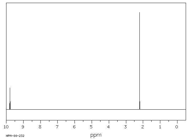
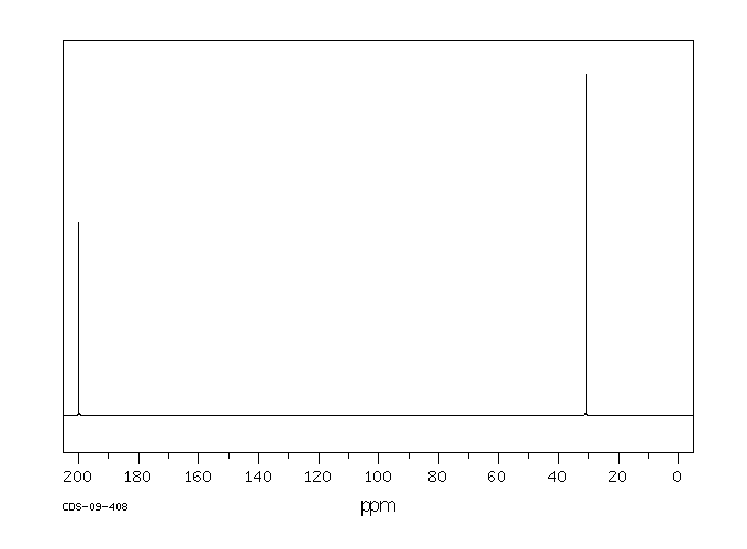
Acetaldehyde appears as a clear colorless liquid with a pungent choking odor. Flash point -36°F. Boiling point 69°F. Density 6.5 lb / gal. Vapors are heaver than air and irritate the mucous membranes and especially the eyes. Used to make other chemicals.
颜色/状态:
Volatile liquid or gas
气味:
Pungent, fruity odor
味道:
Tart taste (fruits containing acetaldehyde before ripening)
Decomposes above 400 °C to form ... methane & carbon monoxide.
粘度:
0.253 mPa s at 9.5 °C; 0.21 mPa s at 20 °C
燃烧热:
-1168.79 kJ/mol (liquid at constant pressure)
汽化热:
25.73 kJ/mol at 20.2 °C
表面张力:
21.2 mN/m at 20 °C (1.0 mN/m = 1.0 dyn/cm)
电离电位:
10.22 eV
聚合:
The substance may polymerize under the influence of acids, alkaline materials, such as sodium hydroxide, in the presence of trace metals (iron) with fire or explosion hazard. (From table)
气味阈值:
Recognition in air= 2.1x10⁻¹ ppm (chemically pure)
Alcohol abuse is one of the major causes of liver fibrosis worldwide. Although the pathogenesis of liver fibrosis is a very complex phenomenon involving different molecular and biological mechanisms, several lines of evidence established that the first ethanol metabolite, acetaldehyde, plays a key role in the onset and maintenance of the fibrogenetic process. This review briefly summarizes the molecular mechanisms underlying acetaldehyde pro-fibrogenic effects. Alcoholic Liver Disease (ALD) has a complex pathogenesis, in which acetaldehyde (AcCHO), the major ethanol metabolite, plays a central role. Ethanol is mainly metabolized in the liver by two oxidative pathways. In the first one ethanol is oxidized to acetaldehyde by the cytoplasmic alcohol dehydrogenase enzyme (ADH), acetaldehyde is then oxidized to acetic acid by the mitochondrial acetaldehyde dehydrogenase (ALDH). The second pathway is inducible and involves the microsomal ethanol-oxidizing system (MEOS), in which the oxidation of ethanol to acetaldehyde and acetic acid also leads to generation of reactive oxygen species (ROS). Chronic ethanol consumption significantly inhibits mitochondrial ALDH activity while the rate of ethanol oxidation to acetaldehyde is even enhanced, resulting in a striking increase of tissue and plasma acetaldehyde levels ... This review will focus on the molecular mechanisms by which acetaldehyde promote liver fibrosis.
It is known that drinking alcohol can lead to reproductive problems in women. In this study, we analyzed the possibility that part of those effects were mediated through alterations of ovarian function related to ethanol oxidation to acetaldehyde occurring in situ. Biotransformation in the rat ovary cytosolic fraction was partially inhibited by allopurinol, suggesting the participation of xanthine oxidoreductase in the process. Microsomal pathway was of enzymatic nature, requiring nicotinamide adenine dinucleotide phosphate-oxidase (NADPH), sensitive to oxygen and significantly inhibited by sodium diethyldithiocarbamate, 4-methylpyrazole and diphenyleneiodonium. Aldehyde dehydrogenase activity was detected by histochemistry in the ovarian tissue, in the strome surrounding the follicle while no alcohol dehydrogenase was detected. However, biochemical determination of alcohol dehydrogenase and aldehyde dehydrogenase activities in rat ovarian tissue revealed the presence of some activity of both enzymes but significantly lower than those found in the liver. By repetitive exposure of animals to ethanol, the microsomal metabolism to acetaldehyde was increased but not in the case of the cytosolic fraction. In these animals, t-butylhydroperoxyde-promoted chemiluminiscence was increased in comparison to control, revealing an increased susceptibility to oxidative stress due to alcohol drinking. Ultrastructure of ovarian tissue from rats exposed chronically to alcohol revealed alterations at the level of the granulosa; theca interna and pellucida zones. In the secondary follicle, alterations consisted of marked condensation of chromatin attached to the nuclear inner membrane. Intense dilatation of the outer perinuclear space could be observed. There was a marked dilatation of the rough endoplasmic reticulum accompanied of significant detachment of ribosomes from their membranes. Mitochondria appeared swollen. In the zona pellucida, most of the cell processes from oocyte and corona radiata cells were absent or broken totally or in part. Results suggest that in the rat ovary, metabolism of ethanol to acetaldehyde may play a role in alcohol effects on female reproductive function.
The biosynthesis of androgens requires multiple steps and during the conversion of pregnenolone to 17 alpha-hydroxypregnenolone and dehydroepiandrosterone (DHEA) by CYP17a1. Acetaldehyde is potentially formed as a by-product in theca cells during antral follicular development. In this study, acetaldehyde level was significantly increased after eCG stimulation and reached a maximum level at 36-hr post-eCG. By 48 hr, the level of acetaldehyde decreased in association with the induction of aldehyde dehydrogenase (ALDH) type 1 family members. When immature mice were co-injected with the ALDH inhibitor, cyanamide, and eCG, the expression of genes involved in the differentiations of granulosa cells was suppressed and the number of ovulated oocytes was reduced. The in vitro studies showed that ALDH inhibitors prevented FSH-induced granulosa cell differentiation. These results indicate that acetaldehyde is generated as a by-product during steroidogenesis and can exert toxic effects to impair the differentiation of granulosa cells, reduce ovulation and decrease oocyte quality.
... The effects of ethyl alcohol are indicative of effects of acetaldehyde, because it is the major metabolite of ethyl alcohol. /Acetaldehyde is/ ... the principal metabolic buildup product of disulfiram therapy.
Acetaldehyde has known human metabolites that include Acetic Acid.
来源:NORMAN Suspect List Exchange
毒理性
毒性总结
乙醛是一种无色挥发性液体,具有刺鼻的窒息性气味。目前在美国没有注册为当前的农药使用,但批准的农药用途可能会定期更改,因此必须咨询联邦、州和地方当局以获取当前批准的用途。乙醛用作生产纤维素醋酸酯、醋酸乙烯树脂、醋酸酯、合成吡啶衍生物和对苯二甲酸的中间体。其他用途包括:在镜子镀银中;在制革中,作为酒精的变性剂;在燃料混合物中;作为明胶纤维的硬化剂;在胶水和酪蛋白产品中;作为鱼类和水果的防腐剂;在造纸工业中;作为合成香料剂;以及化妆品的制造中。乙醛已被确认为在液压破碎液中常用的物质。人类暴露和毒性:乙醛是人类代谢的中间体。它已被发现在食物、饮料和香烟烟雾中。到目前为止,一般人群中暴露于乙醛的主要来源是通过乙醇的代谢。在某些制造行业和酒精发酵期间,工人可能会暴露。在人类肝脏和其他组织中已经鉴定出乙醛脱氢酶(ALDH)的几种同工酶形式。线粒体ALDH存在多态性。对于线粒体ADLH相应基因中的点突变呈纯合子或杂合子的受试者,这种酶的活性较低,代谢乙醛缓慢,对乙醇酒精不耐受。人类肾小管中也有一些乙醛的代谢;肝脏是最重要的代谢场所。在涉及人类志愿者的有限研究中,短期暴露后乙醛对眼睛和上呼吸道有轻微刺激性。将5%的乙醛以20.6-82.4 mg/min的速度静脉输注到正常人类受试者体内,持续36分钟,导致心率、通气和死腔增加,肺泡二氧化碳水平降低。这些症状在质量和数量上与接受双硫仑(Antabuse)治疗的受试者饮用乙醇后看到的症状相似,双硫仑是一种已知的ALDH抑制剂。大量服用可能导致呼吸麻痹而死亡。慢性中毒的症状类似于慢性酒精中毒。乙醛被认为是诱导乙醇酒精相关肝损伤、面部潮红和发育效应的假定毒性代谢物。来自已知酒精成瘾者的 human lymphocytes (人淋巴细胞)暴露于0.02 mg/mL和0.04 mg/mL的乙醛浓度。结果表明,在这两个浓度下都发生了染色体畸变。在人类酒精滥用者的粒细胞和淋巴细胞中观察到了乙醛-DNA加合物。动物研究:在重复剂量研究中,无论是口服还是吸入途径,相对较低浓度的毒性效应仅限于初始接触部位。在一项研究中,大鼠通过饮用水给予乙醛,效果仅限于前胃的轻微局部角化过度。在通过吸入暴露于乙醛的仓鼠中,观察到呼吸道的效应,同时在喉部观察到退行性变化。在更高的浓度下,注意到呼吸道上皮和喉部的退行性变化。大鼠吸入乙醛后,乙醛会分布到血液、肝脏、肾脏、脾脏、心脏和其他组织中。在母体ip注射乙醛(小鼠)和母体暴露于乙醇(小鼠和大鼠)后,在胚胎中检测到低水平。怀孕的大鼠和小鼠经parenteral暴露于乙醛后,诱导胎儿畸形。乙醛在体外是致突变的,诱导哺乳动物细胞在无外源代谢激活的情况下发生基因突变、断裂效应和姐妹染色单体交换(SCEs)。在Salmonella中没有观察到阴性结果。ip注射后,乙醛诱导了中国仓鼠和小鼠骨髓中的SCEs。这种化学物质ip给药并没有增加早期小鼠精原细胞中微核的频率。在大鼠和仓鼠吸入乙醛的研究中,注意到肿瘤发生率增加。在大鼠中,与剂量有关的鼻腺癌和鳞状细胞癌增加。在仓鼠中,鼻腔和喉部癌症的增加不显著。乙醛分布到脑间质液,但没有分布到脑细胞,这是在ip注射乙醇后得到证实的。在狒狒消耗后,乙醛被红细胞摄取。口服给药后,几乎没有未改变的乙醛在尿液中排出。乙醛代谢的主要途径是通过氧化成醋酸。生态毒性研究:乙醛(0.1%或1.0%,2小时)诱导了影响秀丽隐杆线虫产卵系统的基因突变。
IDENTIFICATION AND USE: Acetaldehyde is a colorless volatile liquid with a pungent suffocating odor. It is not registered for current pesticide use in the U.S., but approved pesticide uses may change periodically and so federal, state and local authorities must be consulted for currently approved use. Acetaldehyde used as an intermediate in the production of cellulose acetate, vinyl acetate resins, acetate esters, synthetic pyridine derivatives, and terephthalic acid. Other uses include: in the silvering of mirrors; in leather tanning, as a denaturant for alcohol; in fuel mixtures; as a hardener for gelatin fibers; in glue and casein products; as a preservative for fish and fruit; in the paper industry; as a synthetic flavoring agent; and in the manufacture of cosmetics. Acetaldehyde has been identified as being commonly used in hydraulic fracturing fluids. HUMAN EXPOSURE AND TOXICITY: Acetaldehyde is a metabolic intermediate in humans. It has been identified in food, beverages and cigarette smoke. By far, the main source of exposure to acetaldehyde in the general population is through metabolism of ethanol. Workers may be exposed in some manufacturing industries and during alcohol fermentation. Several isoenzymic forms of acetaldehyde dehydrogenase (ALDH) have been identified in the human liver and other tissues. There is polymorphism for mitochondrial ALDH. Subjects that are homozygous or heterozygous for a point mutation in the mitochondrial ADLH corresponding gene have low activity of this enzyme, metabolize acetaldehyde slowly and are intolerant of ethanol alcohol. There is some metabolism of acetaldehyde in human renal tubules; the liver is the most important metabolic site. Limited studies involving human volunteers have shown that acetaldehyde was mildly irritating to the eyes and upper respiratory tract following short term exposures. Intravenous infusion of 5% acetaldehyde at a rate of 20.6- 82.4 mg/min for up to 36 min into normal human subjects caused an increase in heart rate, ventilation and dead space, and a decrease in alveolar carbon dioxide levels. These symptoms are qualitatively and quantitatively similar to those seen after ethanol intake in subjects previously treated with disulfiram (Antabuse), a known inhibitor of ALDH. Large doses may cause death by respiratory paralysis. Symptoms of chronic intoxication resemble those of chronic alcoholism. Acetaldehyde has been implicated as the putatively toxic metabolite in the induction of ethanol alcohol associated liver damage, facial flushing and developmental effects. Human lymphocytes (from known alcoholics) were exposed to acetaldehyde concn of 0.02 mg/mL and 0.04 mg/mL. Results indicate that chromosomal aberrations occurred at both concentrations. Acetaldehyde-DNA adducts have been observed in granulocytes and lymphocytes of human alcohol abusers. ANIMAL STUDIES: In repeated dose studies, both the oral and inhalation routes, toxic effects at relatively low concentrations were limited to the sites of initial contact. In a study where rats were administered acetaldehyde in their drinking water effects were limited to slight focal hyperkeratosis of the forestomach. Respiratory effects were noted in hamsters exposed to acetaldehyde by inhalation, degenerative changes were observed in the trachea. Degenerative changes in respiratory epithelium and larynx were noted at higher concentrations. Following inhalation by rats, acetaldehyde is distributed to the blood, liver, kidney, spleen, heart and other tissues. Low levels were detected in embryos after maternal ip injection of acetaldehyde (mouse) and following maternal exposure to ethanol (mouse and rat). Parenteral exposure of pregnant rats and mice to acetaldehyde induced fetal malformations. Acetaldehyde is genotoxic in vitro, inducing gene mutations, clastogenic effects and sister chromatid exchanges (SCEs) in mammalian cells in the absence of exogenous metabolic activation. Negative results were noted with Salmonella. Following ip injection, acetaldehyde induced SCEs in the bone marrow of Chinese hamsters and mice. This chemical administered ip did not increase the frequency of micronuclei in early mouse spermatids. Increased incidences of tumors have been noted in inhalation studies on rats and hamsters exposed to acetaldehyde. In rats, there were dose related increases in nasal adenocarcinomas and squamous cell carcinomas. In hamsters, increases in nasal and laryngeal carcinomas were non-significant. Distribution of acetaldehyde to brain interstitial fluid, but not to brain cells has been demonstrated following ip injection of ethanol. Acetaldehyde is taken up by red blood cells following consumption in baboons. Following oral administration, virtually no unchanged acetaldehyde is excreted in the urine. The major pathway for the metabolism of acetaldehyde is by oxidation to acetate. ECOTOXICITY STUDIES: Acetaldehyde (0.1% or 1.0% for 2 hr) induced mutations in genes that affect the egg-laying system of Caenorhabditis elegans.
Evaluation: There is inadequate evidence in humans for the carcinogenicity of acetaldehyde. There is sufficient evidence in experimental animals for the carcinogenicity of acetaldehyde. Overall evaluation: Acetaldehyde is possibly carcinogenic to humans (Group 2B).
Penetration of acetaldehyde into cerebrospinal fluid (CSF) was studied in healthy human volunteers during calcium carbimide-ethanol interaction. CSF was sampled via lumbar puncture and blood from a cubital vein. CSF and blood acetaldehyde concentrations varied from 1 to 41 and from 22 to 138 mumol/L, respectively. The results indicate that acetaldehyde penetrates the human blood-liquor barrier. Computer analysis of electroencephalograms (EEGs) recorded during the interaction showed reduction in alpha activity with a concomitant increase in delta activity. The changes were similar to those previously observed during 'normal' ethanol intoxication.
Hepatic and blood acetaldehyde concentrations during ethanol oxidation were determined in C57 and DBA mice. Liver acetaldehyde was determined with the perchloric acid-thiourea method (no artifactual acetaldehyde formation). Levels ranging from 5 to 118 nmole/g were observed. At ethanol concentrations below 50-60 umole/g, liver acetaldehyde concentrations were higher in DBA compared with C57 mice. A positive correlation was found between the ethanol and acetaldehyde concentration, when ethanol concentration was below 25 (DBA) or 70 umole/g (C57). At higher ethanol concentrations the correlations tended to become negative. Hemolysis causes artifactual formation of acetaldehyde when blood acetaldehyde is determined using thiourea or semicarbazide methods. The magnitude of the artifactually formed acetaldehyde was of such order that no conclusions regarding the existence of true in vivo blood acetaldehyde concentrations could be drawn.
The effect of ethanol on tissue distribution of acetaldehyde following exposure to acetaldehyde was studied in rats. Male Sprague-Dawley rats were exposed to 20 mM acetaldehyde vapor for 1 hr. They were also administered 0 or 3 g/kg ethanol intragastrically. Blood samples were taken at selected times after acetaldehyde exposure was discontinued and analyzed for acetaldehyde. Other animals were killed immediately after acetaldehyde exposure or 3 hr after ethanol and the liver, blood, kidney, spleen, heart muscle, and skeletal were taken for acetaldehyde analysis. Acetaldehyde disappeared rapidly from the blood with a half-life of 3.1 min. After acetaldehyde inhalation, peripheral blood had the highest acetaldehyde content; other tissue concn were similar except for the liver which had a much lower acetaldehyde content. Acetaldehyde concn after ethanol admin were higher in the liver and blood than in other tissues. ... Blood acetaldehyde is not taken up by hepatocytes to any appreciable extent or hepatic acetaldehyde metabolism is very rapid. ... Inhalation exposure is a viable model for studying acetaldehyde toxicity.
Plant Growth Regulator Daminozide Is a Selective Inhibitor of Human KDM2/7 Histone Demethylases
作者:Nathan R. Rose、Esther C. Y. Woon、Anthony Tumber、Louise J. Walport、Rasheduzzaman Chowdhury、Xuan Shirley Li、Oliver N. F. King、Clarisse Lejeune、Stanley S. Ng、Tobias Krojer、Mun Chiang Chan、Anna M. Rydzik、Richard J. Hopkinson、Ka Hing Che、Michelle Daniel、Claire Strain-Damerell、Carina Gileadi、Grazyna Kochan、Ivanhoe K. H. Leung、James Dunford、Kar Kheng Yeoh、Peter J. Ratcliffe、Nicola Burgess-Brown、Frank von Delft、Susanne Muller、Brian Marsden、Paul E. Brennan、Michael A. McDonough、Udo Oppermann、Robert J. Klose、Christopher J. Schofield、Akane Kawamura
DOI:10.1021/jm300677j
日期:2012.7.26
N-demethylation of Nε-methyl lysine residues in histones and are current therapeutic targets. A set of human 2-oxoglutarate analogues were screened using a unified assay platform for JmjC demethylases and related oxygenases. Results led to the finding that daminozide (N-(dimethylamino)succinamic acid, 160 Da), a plant growth regulator, selectivelyinhibits the KDM2/7 JmjC subfamily. Kinetic and crystallographic
C5 설폰 화합물, 그의 제조방법 및, 이를 이용한 크로세틴 디니트릴 제조방법 및 그의 용도
申请人:Myongji University Industry and Academia Cooperation Foundation 명지대학교 산학협력단(220050139720) BRN ▼135-82-11060
公开号:KR20150115997A
公开(公告)日:2015-10-15
본 발명은 신규의 C5 설폰 화합물, 그 제조방법 및 이를 이용한 크로세틴 디니트릴 제조방법에 관한 것으로, 더욱 상세하게는 하이드라존 보호기를 갖는 신규의 C5 설폰 화합물, 그 제조방법과 이를 이용하여 크로세틴 디니트릴 화합물을 효율적으로 제조할 수 있는 방법에 관한 것이다. 본 발명에 따른 신규의 설폰 화합물은 안정하고, 결정성이 좋아 분리 정제가 용이하고, 이중 결합 형성시 E-구조 형성이 용이하다. 또한 상기 설폰 화합물을 이용하여 크로세틴 디니트릴을 합성하는 경우 결합 반응의 부산물인 설핀 산의 제거가 용이하기 때문에 순수한 최종 생성물을 효율적으로 얻을 수 있다. 본 발명에 따른 크로세틴 디니트릴 화합물은 카로틴 화합물의 일종으로 양 말단에 니트릴기를 함유하여 다양한 반응성을 기대할 수 있다. 또한 양 말단에 포함된 질소원자는 금속에 대한 친화력이 우수하기 때문에 금속 표면에 자기 조립이 가능하다. 아울러 카로틴의 일반적인 항산화 기능을 나타낼 수 있고 유기 분자 도선 등 전기 전자 재료로서도 사용이 가능하다.
Phenanthroline-7-one derivatives and their therapeutic uses
申请人:Laboratoire L. Lafon
公开号:US06809096B1
公开(公告)日:2004-10-26
A pharmaceutical composition including an efficient amount of a compound selected among the compounds of formulae (I) and (Ia). The compounds have interesting cytotoxic properties leading to a therapeutic use as antitumoral medicines.
A Novel, One-Pot Synthesis of α-<i>C</i>-Cyanohydrazines in the Presence of Lithium Perchlorate/Diethylether Solution (5.0 M)
作者:Akbar Heydari、Robabe Baharfar、Mohsen Rezaie、Saied M. Aslanzadeh
DOI:10.1246/cl.2002.368
日期:2002.3
Condensation of N,N-dimethylhydrazine, an aldehyde in lithium perchlorate/diethylether solution (5.0 M) gave N,N-dimethylhydrazone, which were treated with trimethylsilyl-cyanide to afford α-C-cyanohydrazine. These compounds are important precursors of nitrogen-substituted reagents.
[EN] THIOPHENE DERIVATIVES FOR THE TREATMENT OF DISORDERS CAUSED BY IGE<br/>[FR] DÉRIVÉS DE THIOPHÈNE POUR LE TRAITEMENT DE TROUBLES PROVOQUÉS PAR IGE
申请人:UCB BIOPHARMA SRL
公开号:WO2019243550A1
公开(公告)日:2019-12-26
Thiophene derivatives of formula (I) and a pharmaceutically acceptable salt thereof are provided. These compounds have utility for the treatment or prevention of disorders caused by IgE, such as allergy, type 1 hypersensitivity or familiar sinus inflammation.