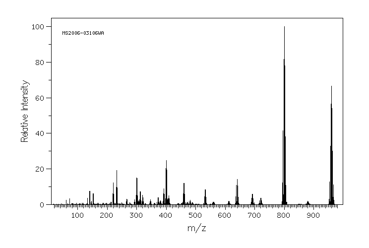
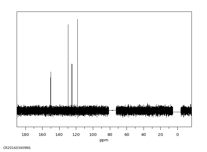
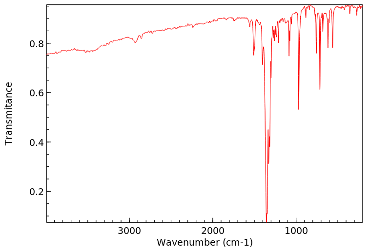
... The unconjugated phenolic fractions isolated from the feces were examined by gas chromatography/mass spectrometry (GC/MS). Methoxyhydroxylated penta- to heptabrominated diphenyl ethers were identified. ... The methoxy and hydroxy substituents were on the same phenyl ring when both were present. In addition, trace amounts of debrominated metabolites and nonaBDEs were also found in the feces and bile, indicating debromination may have been the first step in decaBDE metabolism. A small proportion of monohydroxylated metabolites was found in tissues and feces, indicating a role for reductive dehalogenation followed by an oxidation step or direct oxidative dehalogenation reactions.
About 20% of the decaBDE-derived radioactivity in urine from noncannulated rats was protein bound at 72 hours (compared to >73% not associated with protein). Eighteen percent of the bound material was bound to albumin, a serum protein with the ability to bind (nonspecific protein binding) short chain fatty acids. In cannulated rats, 18.2% of the decaBDE-derived radioactivity was unbound, and all the remaining 68.3% was associated with albumin. Two polar metabolites were noted but not identified. Under the assumption that the bound materials are less polar than the unbound materials, this observation supports the concept that a substantial portion, but not all, of the metabolites in the fecal matter originate from the bile. About 90% of the biliary radiolabel was associated with an unidentified 79-kDa protein, with the percent of bound label decreasing from 94 to 87% in bile samples pooled at 1-24 hours, 24-48 hours, and 48-72 hours. Approximately 17% of the protein-bound biliary radioactivity collected over the first 24 hours was parent compound (and the remainder was unidentified metabolites); no parent compound was detected at 48 and 72 hours. The percent of total bound label in the bile samples also declined over time. None of the label in the bile was found to be unbound.
The metabolism of unlabeled decaBDE /was evaluated/ in male rats after gavage or i.v. injection. Blood samples were collected at specific intervals for up to 6 days. Pooled plasma samples from all 6 days were extracted and decaBDE and its metabolites were quantified. Analysis of the pooled samples indicated that the major neutral compound in the plasma was unmodified decaBDE with trace amounts of three nonaBDEs. Thirteen phenolic metabolites were determined in the plasma of both the orally and i.v. dosed rats, but only three phenolic metabolites were present in sufficiently high concentration for further analysis. These metabolites were characterized as a hydroxy-octaBDE, an hydroxy-nonaBDE, and an hydroxy/methoxy hexaBDE. The relative amount of each metabolite recovered was not reported, but the concentration of phenolic radioactivity in the plasma collected 3 and 7 days after oral gavage was four times higher than that of the neutral compounds (i.e., the parent or debrominated decaBDE). The authors indicated that reductive debromination may be the first step in the metabolic pathway of decaBDE, followed by oxidation to form phenolic metabolites. It was also suggested that the hydroxy/methoxy metabolites were probably formed via an arene oxide hydrolyzed to a dihydrodiol and further rearomatized followed by a methylation reaction.
DecaBDE metabolism /was studied/ in rats given a single oral dose by gavage. Results indicated that in noncannulated rats about 90% of the dose was excreted in the feces within 3 days after a single oral dose of 14C-labeled decaBDE and the majority of this radioactivity (65%) represented decaBDE metabolites. Measurement of bile radioactivity indicated that close to 10% of the total dose was excreted in the bile during the same period, with almost all of the excreted dose in bile in the form of metabolites. Analysis of radiolabeled materials from tissues of these rats at day 3 after decaBDE administration revealed that 42% of the radioactivity in the liver represented solvent extractable lipid-bound metabolites and 30% was in the form of extractable unconjugated metabolites (4 and 26% of which were hydroxylated and neutral metabolites, respectively). Twenty-seven percent of the radiolabel could not be extracted and was tissue bound. Only 1% of the extractable material was water soluble. A larger percentage (61%) of the radioactivity in the small intestine wall was tissue bound. Lipid-bound metabolites accounted for 7% of the label, water-soluble compounds for 11%, and unbound parent or metabolites for 20%. The percentage of the radiolabel found as water-soluble compounds in the intestinal wall was 10 times greater than the water-soluble metabolites in the liver, providing some support for the hypothesis that oxidative metabolism can occur in the intestinal mucosa. Most of the radioactivity (71-80%) in the lung, adipose tissue, and kidney was unbound parent or metabolites; 15-21% represented lipid-bound metabolites, and 1.5-8% was tissue bound. Formation of adducts was indicative of covalent and/or noncovalent interactions with cellular macromolecules.
Polybrominated biphenyls can be absorbed through oral, inhalation, and dermal routes. Once in the body they distribute throughout and bioaccumulate in the blood, breast milk, and adipose tissue. The extent of PBDE metabolism depends on the degree of bromination. Metabolism is believed to involve debromination and methylation, resulting in phenolic metabolites. Metabolized and unmetabolized PDBE compounds are excreted mainly in the faeces. (L628, L881)
IDENTIFICATION AND USE: Decabromodiphenyl ether (deca-BDE) is used as a flame retardant. It is mostly used in applications in the plastics and textile industries. It is an additive flame retardant, i.e. it is physically combined with the material being treated rather than chemically combined (as in reactive flame retardants). HUMAN EXPOSURE AND TOXICITY: Deca-BDE did not produce skin sensitization in human subjects. A health assessment of workers exposed for at least 6 weeks to polybromodiphenyls and polybromodiphenyl oxides, including deca-BDE, during manufacture revealed a higher than normal prevalence of primary hypothyroidism with elevated serum concentrations of thyrotropin and low or borderline-low, serum T4 and free thyroxine indexes in 4 of the 35 occupationally exposed vs. 0 of the 89 control subjects. A significant reduction in sensory and fibula motor velocities was also observed. This primary hypothyroidism was partially reversible in 1 of the 3 workers re-evaluated one year after the initial study. The 2 other workers reassessed still exhibited low free thyroxine indexes and high thyrotrophin values. Significant correlation was seen between length of employment and concentrations of follicle stimulating hormone in workers exposed to deca-BDE An abnormal follicle stimulating hormone value was found in only one worker. A testicular cyst was found in one exposed worker, and epididymal nodules in two others. No testicular or epididymal nodules were seen among comparisons. In other studies deca-BDE was detected in human serum, milk, and sperm. Deca-BDE induced DNA damage in human neuroblastoma cells. ANIMAL STUDIES: Deca-BDE caused no dermal response, and did not cause primary eye irritation in rabbits. Deca-BDE is poorly absorbed and does not easily penetrate the cell wall. Its acute and chronic toxicities are relatively low, with the liver and the thyroid as the primary targets, though there is some evidence of carcinogenicity. In NTP 2-year feeding studies, there was some evidence of carcinogenicity for male and female rats as shown by increased incidences of neoplastic nodules of the liver in low dose (25,000 ppm) males and high dose (50,000 ppm) groups of each sex. There was equivocal evidence of carcinogenicity for male mice as shown by increased incidences of hepatocellular adenomas or carcinomas (combined) in the low dose group and of thyroid gland follicular cell adenomas or carcinomas (combined) in both dosed groups. There was no evidence of carcinogenicity for female mice receiving 25,000 or 50,000 ppm in the diet. Several non-neoplastic lesions were observed at increased incidences, the most notable being thyroid gland follicular cell hyperplasia in male mice. Several animal studies have indicated that deca-BDE may cause developmental neurotoxicity, affecting motor and cognitive domains. Deca-BDE exposure during pregnancy and lactation impaired immune function in rats. Several in vivo and in vitro studies have also demonstrated effects of deca-BDE on thyroid hormone homeostasis. Deca-BDE was not genotoxic in Salmonella typhimurium TA98-100-1535-1537 and Escherichia coli WP2 uvr with or without activation. It was also not mutagenic in the mouse lymphoma L5178y/TK + or - assay in the presence or absence of metabolic activation. Tests for cytogenetic effects in Chinese hamster ovary cells indicated that this chemical does not cause chromosomal aberrations or sister chromatid exchanges either in the presence or absence of activation. ECOTOXICITY STUDIES: Deca-BDE was found in tissues of birds, mammals, and in aquatic species. Metabolism via debromination appears to be a major degradation route of Deca-PBE in juvenile sole in comparison to biotransformation into hydroxylated metabolites. In plants Deca-BDE exposure could cause oxidative stress and damage.
Like other halogenated aromatic hydrocarbons, polybrominated diphenyl ethers bind to the cellular aryl hydrocarbon receptor (AhR), which regulates the synthesis of a variety of proteins. Activation of the AhR induces a number of enzymes, including cytochrome P-450-dependent monooxygenases of the CYP1A and CYP2B families, UDP-glucuronosyltransferase, and ethoxyresorufin-o-deethylase. PBDEs are also believed to disrupt the production, transport, and disposition of thyroid hormones. One mechanism of this involves metabolites ot PDBEs competing with thyroxine to bind to transthyretin, decreasing serum thyroid hormone levels. This change in thyroid hormone levels has been linked to both thyroid toxicity and neurobehavioral alterations. Certain PDBEs and their metabolites are also endocrine disruptors and may act as agonists at the estrogen receptors or antagonists at the androgen and progesterone receptors. (L628, A262)
Evaluation: No epidemiological data relevant to the carcinogenicity of decabromodiphenyl oxide. There is limited evidence in experimental animals for the carcinogenicity of decabromodiphenyl oxide. Overall evaluation: Decabromodiphenyl oxide is not classifiable as to its carcinogenicity to humans (Group 3).
CLASSIFICATION: C; possible human carcinogen. BASIS FOR CLASSIFICATION: Based on no human data and limited evidence of carcinogenicity in animals; namely, significantly increased incidences of neoplastic liver nodules in male and female rats and increased incidences of hepatocellular adenomas or carcinomas (combined) in male mice. HUMAN CARCINOGENICITY DATA: None. ANIMAL CARCINOGENICITY DATA: Limited. /Based on former classification system/
Several studies have demonstrated that decabromodiphenyl oxide (DBDPO) is very poorly absorbed in rats following oral exposure, generally <1% of the amount ingested.
Studies with (14)C labeled decabromodiphenyl oxide administered orally to Sprague Dawley rats indicate that more than 99% of the administered label was excreted in feces within 2 days following administration. An analysis of bromine in tissues following long term exposure in diets that provided 0.1 mg/kg per day to rats indicated a slight increase in bromine content in liver and adipose tissue at 90 days but no significant increase following 12 months of exposure. A significant increase in the bromine content of adipose, but no other tissues, was observed following a similiar dose of decabromodiphenyl oxide for 2 years but not at lower doses. There was no indication as to whether the failure of decabromodiphenyl oxide to accumulate in tissues was due to lack of absorption from the gastrointestinal tract or rapid metabolism and clearance.
Retention of decabromodiphenyl oxide in the brain of neonatal mice /had recently reported/. A single oral dose of 14(C)-labelled decabromodiphenyl oxide (purity >98%) was given on postnatal day 3, 10 or 19 to neonatal NMRI mice. Two litters in each age categories were given 1.5 14(C) MBq/kg body weight. The radioactivity in the brain was determined after 24 hours or 7 days after dosing in each of the two litters from the three different age categories. The results of the study showed that 14(C) was taken up into the brain, but there were differences in the amount of radioactivity found in the different age mice. The mice exposed on postnatal day 3 or 10 had around 4% of the total administered dose of 14(C) in the brain at 24-hours after dosing, whereas only 0.6 % of the total administered dose was found at 24-hours in the brains of mice dosed on postnatal day 19. At day-7 after administration the amount of radioactivity in the brain had increased by around a factor of 2 in the mice exposed on postnatal days 3 or 10, whereas no noticeable change in the amount of radioactivity present had occurred in brains of the mice dosed on postnatal day.