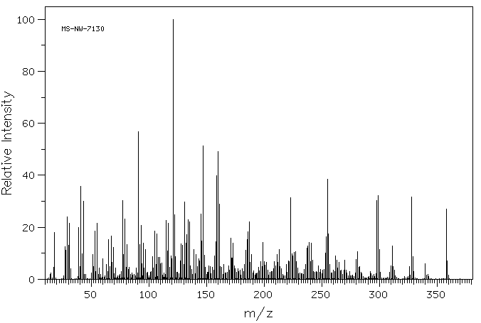
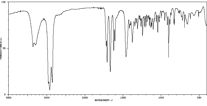
Prednisone is metabolized to 17α,21-dihydroxy-pregnan-1,4,6-trien-3,11,30-trione (M-XVII), 20α-dihydro-prednisone (M-V), 6βhydroxy-prednisone (M-XII), 6α-hydroxy-prednisone (M-XIII), or 20β-dihydro-prednisone (M-IV). 20β-dihydro-prednisone is metabolized to 17α,20ξ,21-trihydroxy-5ξ-pregn-1-en-3,11-dione(M-XVIII). Prednison is reversibly metabolized to [prednisolone]. Prednisolone is metabolized to Δ6-prednisolone (M-XI), 20α-dihydro-prednisolone (M-III), 20β-dihydro-prednisolone (M-II), 6αhydroxy-prednisolone (M-VII), or 6βhydroxy-prednisolone(M-VI). 6αhydroxy-prednisolone is metabolized to 6α,11β,17α,20β,21-pentahydroxypregnan-1,4-diene-3-one (M-X). 6βhydroxy-prednisolone is metabolized to 6β,11β,17α,20β,21-pentahydroxypregnan-1,4-diene-3-one (M-VIII), 6β,11β,17α,20α,21-pentahydroxypregnan-1,4-diene-3-one (M-IX), and 6β,11β,17α,21-tetrahydroxy-5ξ-pregn-1-en-3,20-dione (M-XIV). MVIII is metabolized to 6β,11β,17α,20β,21-pentahydroxy-5ξ-pregn-1-en-3-one (M-XV) and then to MXIV, while MIX is metabolized to 6β,11β,17α,20α,21-pentahydroxy-5ξ-pregn-1-en-3-one (M-XVI) and then to MXIV. These metabolites and their glucuronide conjugates are excreted predominantly in the urine.
In one study after an oral dose of prednisone, the plasma prednisolone concentration peaked between 60 and 120 min and then declined exponentially. After rapid iv injection of steroid, the plasma prednisolone concentration peaked within 10 to 20 min. An initial rapid distribution phase succeeded by a slower decay phase was expressed by a biphasic exponential disappearance curve of the plasma prednisolone concentration versus time. Plasma prednisolone concentrations achieved with an oral dose of prednisone were in the same range as those obtained during the second phase after iv administration.
Reduction of the 11-oxo to the 11alpha-hydroxyl group by the enzyme 11beta-hydroxydehydrogenase converts prednisone to prednisolone, its biologically active form. This reaction takes place mainly in the liver, and may proceed satisfactorily even in the presence of liver disease
来源:Hazardous Substances Data Bank (HSDB)
代谢
在体外,泼尼松被肝脏、肺和肾脏组织转化为泼尼松龙。相反,泼尼松龙被肾脏组织转化为泼尼松。
In vitro, prednisone is converted to prednisolone by liver, lung and renal tissue. Conversely, prednisolone is converted to prednisone by renal tissue.
... The aim of this work was to evaluate the effects of these corticosteroids on the expression of several forms of cytochromes p450, including p450 1A2, 2D6, 2E1, and 3A, and on cyclosporin A oxidase activity in human liver. For this purpose, human hepatocytes prepared from lobectomies were maintained in culture in a serum-free medium, in collagen-coated dishes, for 96-144 hr, in the absence or presence of 50-100 uM corticosteroids, rifampicin, or dexamethasone. To mimic more closely the current clinical protocol, hepatocyte cultures were also co-treated with corticosteroids and cyclosporin A or ketoconazole (a selective inhibitor of cytochromes p450 3A). Cyclosporin A oxidase activity, intracellular retention of cyclosporin A oxidized metabolites within hepatocytes, accumulation of cytochromes p450 proteins and corresponding messages, and de novo synthesis and half-lives of these cytochromes p450 were measured in parallel in these cultures. Our results, obtained from seven different hepatocyte cultures, showed that 1) dexamethasone and prednisone, but not prednisolone or methylprednisolone, were inducers of cytochrome p450 3A, at the level of protein and mRNA accumulation, as well as of cyclosporin A oxidase activity, known to be predominantly catalyzed by these cytochromes p450; 2) although corticosteroids are known to be metabolized in human liver, notably by cytochrome p450 3A, partial or total inhibition of this cytochromes p450 by cyclosporin or ketoconazole, respectively, did not affect the inducing efficiency of these molecules; 3) corticosteroids did not affect the half-life of cytochrome p450 3A or the accumulation of other forms of cytochromes p450, including 1A2, 2D6, and 2E1; 4) chronic treatment of cells with cyclosporin did not affect cytochrome p450 3A accumulation; 5) corticosteroids were all competitive inhibitors of cyclosporin A oxidase in human liver microsomes, with Ki values of 61 + or - 12, 125 + or - 25, 190 + or - 38, and 210 + or - 42 uM for dexamethasone, prednisolone, prednisone, and methylprednisolone, respectively; and 6) chronic treatment of cells with corticosteroids did not influence the excretion of oxidized metabolites of cyclosporin from the cells.
IDENTIFICATION AND USE: Prednisone is white crystalline powder with a persistent bitter after-taste. It is glucocorticoid, anti-inflammatory agent and antineoplastic agent. HUMAN EXPOSURE AND TOXICITY: Prednisone causes profound and varied metabolic effects when used at therapeutic doses, given on a continuous basis. When given in large doses it can induce cardiac complications. In addition, it modifies the body's immune response to diverse stimuli; among the changes that occur are lymphopenia, monocytopenia and suppression of delayed hypersensitivity skin tests. Fluid and electrolyte disturbances may occur, including sodium and fluid retention, which may lead to congestive heart failure and hypertension. With large doses, potassium loss, hypokalemic alkalosis, and increased calcium excretion may occur. Glucocorticoids may cause fetal damage when administered to pregnant women. One retrospective study of 260 women who received pharmacologic dosages of glucocorticoids during pregnancy revealed 2 instances of cleft palate, 8 stillbirths, 1 spontaneous abortion, and 15 premature births. Another study reported 2 cases of cleft palate in 86 births. No chromosomal damage was detected in peripheral lymphocytes of patients treated with 3 mg/kg bw per day prednisone alone for 28 days and then with 0.5-1 mg/kg bw per day for 18-120 months. ANIMAL STUDIES: Carcinogenicity studies have been conducted in rats and mice. Of treated males, 7/20 developed tumors, among which were 3 pituitary tumors and 1 tumor of the breast; 16/18 female rats developed tumors, 8 of the breast, 5 of the pituitary, 2 of the adrenal and 1 of the liver. The overall tumor incidence in females was 1.5-2-fold higher than that in controls. However in mice, the tumor incidence in treated males was 4/19 (21%), with 2 lymphosarcomas and 2 lung tumors, and that in treated females was 8/27 (30%), with 4 lung tumors, 2 lymphosarcomas and 2 uterine tumors. These incidences were not significantly greater than those in controls. Prednisone given to rats from the 11th day of pregnancy until parturition at daily doses of 2.5 or 5 mg was reported to inhibit the growth of the fetal thymus and spleen. Prednisone was not mutagenic in Escherichia coli, and it caused no chromosomal damage when administered to rats. ECOTOXICITY STUDIES: The rotifer Brachionus calyciflorus and two crustaceans, the cladoceran Daphnia magna and the anostracan Thamnocephalus platyurus, were used to perform acute toxicity tests. Chronic toxicity tests have been performed on the alga Pseudokirchneriella subcapitata and the crustacean Ceriodaphnia dubia. The results showed low acute and chronic toxicity of prednisone. Some of the photoproducts had high toxic effects on C. dubia.
Prednisone is a glucocorticoid receptor agonist. It is first metabolized in the liver to its active form, prednisolone. Prednisolone crosses cell membranes and binds with high affinity to specific cytoplasmic receptors. The result includes inhibition of leukocyte infiltration at the site of inflammation, interference in the function of mediators of inflammatory response, suppression of humoral immune responses, and reduction in edema or scar tissue. The antiinflammatory actions of corticosteroids are thought to involve phospholipase A2 inhibitory proteins, lipocortins, which control the biosynthesis of potent mediators of inflammation such as prostaglandins and leukotrienes. Prednisone can stimulate secretion of various components of gastric juice. Suppression of the production of corticotropin may lead to suppression of endogenous corticosteroids. Prednisone has slight mineralocorticoid activity, whereby entry of sodium into cells and loss of intracellular potassium is stimulated. This is particularly evident in the kidney, where rapid ion exchange leads to sodium retention and hypertension.
Oral prednisone has a Tmax of 2 hours, while the delayed-release formulation has a Tmax of 6-6.5 hours. A 5mg dose of prednisone has an AUC of 572mL/min/1.73m2, a 20mg dose of prednisone has an AUC of 1034mL/min/1.73m2, and a 50mg dose of prednisone has an AUC of 2271mL/min/1.73m2. Data regarding the Cmax of prednisone is not readily available.
来源:DrugBank
吸收、分配和排泄
消除途径
泼尼松主要通过尿液以硫酸盐和葡萄糖醛酸苷的形式排出体外。
Prednisone is excreted mainly in the urine as sulfate and glucuronide conjugates.
Data regarding the volume of distribution for prednisone is not readily available. However, a 0.15mg/kg dose of prednisolone has a volume of distribution of 29.3L, while a 0.30mg/kg dose has a volume of distribution of 44.2L.
Data regarding the clearance of prednisone is not readily available. A 5.5µg/h/kg infusion of prednisolone has an average clearance of 0.066±0.12L/h/kg, while a 0.15±0.03L/h/kg infusion has an average clearance of 0.15L/h/kg.
Thirty minutes after iv administration of (3)H-prednisone to a monkey, the concentration of prednisone was highest in the kidney. The drug was also found in the liver, spleen, lung, small intestine, serum and bile. The concentration of prednisolone was highest in the liver. It was also found in the kidney, pancreas, spleen, lung, small intestine, serum and bile.
[EN] ACC INHIBITORS AND USES THEREOF<br/>[FR] INHIBITEURS DE L'ACC ET UTILISATIONS ASSOCIÉES
申请人:GILEAD APOLLO LLC
公开号:WO2017075056A1
公开(公告)日:2017-05-04
The present invention provides compounds I and II useful as inhibitors of Acetyl CoA Carboxylase (ACC), compositions thereof, and methods of using the same.
DISUBSTITUTED TRIFLUOROMETHYL PYRIMIDINONES AND THEIR USE
申请人:BAYER PHARMA AKTIENGESELLSCHAFT
公开号:US20160221965A1
公开(公告)日:2016-08-04
The present application relates to novel 2,5-disubstituted 6-(trifluoromethyl)pyrimidin-4(3H)-one derivatives, to processes for their preparation, to their use alone or in combinations for the treatment and/or prevention of diseases, and to their use for preparing medicaments for the treatment and/or prevention of diseases, in particular for treatment and/or prevention of cardiovascular, renal, inflammatory and fibrotic diseases.
Heterocyclic derivatives for the treatment of cancer and other proliferative diseases
申请人:——
公开号:US20020143182A1
公开(公告)日:2002-10-03
The invention relates to certain heterocyclic compounds useful for the treatment of cancer and other diseases, having the Formula (I):
1
wherein:
(a) m is an integer 0 or 1;
(b) R
12
is an alkyl, a substituted alkyl, a cycloalkyl, a substituted cycloalkyl, a heterocyclic, a substituted heterocyclic, a heteroaryl, a substituted heteroaryl, an aryl or a substituted aryl residue;
(c) Ar
3
is an aryl, a substituted aryl, a heteroaryl or a substituted heteroaryl residue;
(d) Ar
4
is an aryl, a substituted aryl, a heteroaryl or a substituted heteroaryl residue;
(e) R
5
is hydrogen, hydroxy, alkyl or substituted alkyl;
(f) - - - - - represents a bond present or absent; and
(g) W, X, Y and Z are independently or together C(O)—, C(S), S, O, or NH; or a pharmaceutically acceptable salt thereof.
[EN] COMPOUNDS AND COMPOSITIONS COMPRISING CDK INHIBITORS AND METHODS FOR THE TREATMENT OF CANCER<br/>[FR] COMPOSÉS ET COMPOSITIONS COMPRENANT DES INHIBITEURS DES CDK ET MÉTHODES DE TRAITEMENT DU CANCER
申请人:UNIV GEORGIA STATE RES FOUND
公开号:WO2010129858A1
公开(公告)日:2010-11-11
Disclosed herein are compounds suitable for use as antitumor agents, methods for treating cancer wherein the disclosed compounds are used in making a medicament for the treatment of cancer, methods for treating a tumor comprising, administering to a subject a composition comprising one or more of the disclosed cytotoxic agents, and methods for preparing the disclosed antitumor agents.
The present invention provides a cobalamin-drug conjugate suitable for the treatment of tumor related diseases. Cobalamin is indirectly covalently bound to an anti-tumor drug via a cleavable linker and one or more optional spacers. Cobalamin is covalently bound to a first spacer or the cleavable linker via the 5′-OH of the cobalamin ribose ring. The drug is bound to a second spacer of the cleavable linker via an existing or added functional group on the drug. After administration, the conjugate forms a complex with transcobalamin (any of its isoforms). The complex then binds to a receptor on a cell membrane and is taken up into the cell. Once in the cell, an intracellular enzyme cleaves the conjugate thereby releasing the drug. Depending upon the structure of the conjugate, a particular class or type of intracellular enzyme affects the cleavage. Due to the high demand for cobalamin in growing cells, tumor cells typically take up a higher percentage of the conjugate than do normal non-growing cells. The conjugate of the invention advantageously provides a reduced systemic toxicity and enhanced efficacy as compared to a corresponding free drug.