
(6S,2S)-二氨基庚二酸 | 14289-34-0
中文名称
(6S,2S)-二氨基庚二酸
中文别名
——
英文名称
(S,S)-2,6-diaminopimelic acid
英文别名
(2S,6S)-2,6-diaminoheptanedioic acid;(2S,6S)-(+)-2,6-diaminopimelic acid;(2S,6S)-(+)-diaminopimelic acid;(2S,6S)-2,6-diaminopimelic acid;L-2,6-diaminoheptanedioic acid;L,L-2,6-diaminopimelic acid;(2S,6S)-2,6-diaminoheptanedioate;(2S,6S)-2,6-bis(azaniumyl)heptanedioate

CAS
14289-34-0
化学式
C7H14N2O4
mdl
——
分子量
190.199
InChiKey
GMKMEZVLHJARHF-WHFBIAKZSA-N
BEILSTEIN
——
EINECS
——
-
物化性质
-
计算性质
-
ADMET
-
安全信息
-
SDS
-
制备方法与用途
-
上下游信息
-
文献信息
-
表征谱图
-
同类化合物
-
相关功能分类
-
相关结构分类
物化性质
-
沸点:426.7±45.0 °C(Predicted)
-
密度:1.344±0.06 g/cm3(Predicted)
-
熔点:309-312 °C
-
物理描述:Solid
-
碰撞截面:140.02 Ų [M-H]- [CCS Type: DT, Method: stepped-field]
计算性质
-
辛醇/水分配系数(LogP):-5.9
-
重原子数:13
-
可旋转键数:6
-
环数:0.0
-
sp3杂化的碳原子比例:0.71
-
拓扑面积:127
-
氢给体数:4
-
氢受体数:6
安全信息
-
海关编码:2922499990
-
储存条件:| 室温 |
SDS
上下游信息
-
上游原料
中文名称 英文名称 CAS号 化学式 分子量 2,6-二氨基庚二酸 2,6-diaminopimelic acid 583-93-7 C7H14N2O4 190.199 —— (2S,6S)-2,6-Diaminoheptandisaeure-dimethylester 142632-77-7 C9H18N2O4 218.253 -
下游产品
中文名称 英文名称 CAS号 化学式 分子量 —— (2R,6S)-2,6-diaminoheptanedioic acid —— C7H14N2O4 190.199 L-赖氨酸 L-lysine 56-87-1 C6H14N2O2 146.189
反应信息
-
作为反应物:描述:(6S,2S)-二氨基庚二酸 在 potassium hydrogencarbonate 、 三氟乙酸 作用下, 以 乙醇 、 水 为溶剂, 反应 2.0h, 生成 (2S,6S)-2-Amino-6-((S)-2-amino-propionylamino)-heptanedioic acid参考文献:名称:Peptides of 2-aminopimelic acid: antibacterial agents that inhibit diaminopimelic acid biosynthesis摘要:Succinyl-CoA:tetrahydrodipicolinate-N-succinyltransferase is a key enzyme in the biosynthesis of diaminopimelic acid (DAP), a component of the cell wall peptidoglycan of nearly all bacteria. This enzyme converts the cyclic precursor tetrahydrodipicolinic acid (THDPA) to a succinylated acyclic product. L-2-Aminopimelic acid (L-1), an acyclic analogue of THDPA, was found to be a good substrate for this enzyme and was shown to cause a buildup of THDPA in a cell-free enzyme system but was devoid of antibacterial activity. Incorporation of 1 into a di- or tripeptide yielded derivatives that exhibited antibacterial activity against a range of Gram-negative organisms. Of the five peptide derivatives tested, (L-2-aminopimelyl)-L-alanine (6) was the most potent. These peptides were shown to inhibit DAP production in intact resting cells. High levels (30 mM) of 2-aminopimelic acid were achieved in the cytoplasm of bacteria as a result of efficient uptake of the peptide derivatives through specific peptide transport systems followed, presumably, by cleavage by intracellular peptidases. Finally, the antibacterial activity of these peptides could be reversed by DAP or a DAP-containing peptide. These results demonstrate that the peptides containing L-2-aminopimelic acid exert their antibacterial action by inhibition of diaminopimelic acid biosynthesis.DOI:10.1021/jm00151a015
-
作为产物:描述:tert-butyl (2S)-2-{bis[(tert-butoxy)carbonyl]amino}-5-oxopentanoate 在 [(1,2,5,6-r)-1,5-cyclooctadiene][(2S,2'S,5 S,5'S)-1,1'-(1,2-phenylene)bis[2,5-diethylphospholane-κP]]-rhodium(1+) trifluoromethanesulfonate 氢溴酸 、 氢气 、 溶剂黄146 、 1,8-二氮杂双环[5.4.0]十一碳-7-烯 、 苯酚 作用下, 以 甲醇 、 二氯甲烷 为溶剂, 20.0 ℃ 、482.63 kPa 条件下, 反应 44.5h, 生成 (6S,2S)-二氨基庚二酸参考文献:名称:通过不对称氢化高效合成 (2S,6S)- 和内消旋二氨基庚二酸摘要:已成功开发了标题化合物 1 和 2 的有效合成方法。关键步骤是使用 [Rh(I)(COD)-(S,S) 或 -(R,R)-Et-DuPHOS)] + OTf 对脱氢氨基酸 7 进行不对称氢化,以产生具有光学活性的受保护氨基酸高 ee (>95%) 的衍生物 该方法还可用于合成其他异构体和类似物。DOI:10.1055/s-2002-19295
文献信息
-
Synthesis of bis-α,α′-amino acids through diastereoselective bis-alkylations of chiral Ni(ii)-complexes of glycine作者:Jiang Wang、Hong Liu、José Luis Aceña、Daniel Houck、Ryosuke Takeda、Hiroki Moriwaki、Tatsunori Sato、Vadim A. SoloshonokDOI:10.1039/c3ob40594j日期:——conditions for the alkylation reactions have been investigated and the latter proved to be more efficient in terms of stereochemical outcome. In particular, alkylation of the glycine Schiff base Ni(II) complex with 1,3-dibromopropane followed by acid-catalysed hydrolysis of the resulting bis-alkylation product, allowed for the preparation of naturally occurring (2S,6S)-diaminopimelic acid in high overall
-
Synthesis of characteristic Mycobacterium peptidoglycan (PGN) fragments utilizing with chemoenzymatic preparation of meso-diaminopimelic acid (DAP), and their modulation of innate immune responses作者:Qianqian Wang、Yusuke Matsuo、Ambara R. Pradipta、Naohiro Inohara、Yukari Fujimoto、Koichi FukaseDOI:10.1039/c5ob02145f日期:——
Characteristic
Mycobacterium peptidoglycan fragments were comprehensively synthesized and their weaker immunostimulationvia Nod1 and Nod2 was shown.特征性的分子Mycobacterium肽聚糖片段已全面合成,并显示了它们通过Nod1和Nod2较弱的免疫刺激作用。 -
Atomic-Resolution 1.3 Å Crystal Structure, Inhibition by Sulfate, and Molecular Dynamics of the Bacterial Enzyme DapE作者:Matthew Kochert、Boguslaw P. Nocek、Thahani S. Habeeb Mohammad、Elliot Gild、Kaitlyn Lovato、Tahirah K. Heath、Richard C. Holz、Kenneth W. Olsen、Daniel P. BeckerDOI:10.1021/acs.biochem.0c00926日期:2021.3.30We report the atomic-resolution (1.3 Å) X-ray crystal structure of an open conformation of the dapE-encoded N-succinyl-l,l-diaminopimelic acid desuccinylase (DapE, EC 3.5.1.18) from Neisseria meningitidis. This structure [Protein Data Bank (PDB) entry 5UEJ] contains two bound sulfate ions in the active site that mimic the binding of the terminal carboxylates of the N-succinyl-l,l-diaminopimelic acid (l,l-SDAP) substrate. We demonstrated inhibition of DapE by sulfate (IC50 = 13.8 ± 2.8 mM). Comparison with other DapE structures in the PDB demonstrates the flexibility of the interdomain connections of this protein. This high-resolution structure was then utilized as the starting point for targeted molecular dynamics experiments revealing the conformational change from the open form to the closed form that occurs when DapE binds l,l-SDAP and cleaves the amide bond. These simulations demonstrated closure from the open to the closed conformation, the change in RMS throughout the closure, and the independence in the movement of the two DapE subunits. This conformational change occurred in two phases with the catalytic domains moving toward the dimerization domains first, followed by a rotation of catalytic domains relative to the dimerization domains. Although there were no targeting forces, the substrate moved closer to the active site and bound more tightly during the closure event.我们报告了脑膜炎奈瑟菌中由 DAPE 编码的 N-琥珀酰-l,l-二氨基亚庚酸脱琥珀酰化酶(DAPE,EC 3.5.1.18)的开放构象的原子分辨率(1.3 Å)X 射线晶体结构。该结构[蛋白质数据库(PDB)条目 5UEJ]的活性位点含有两个结合的硫酸根离子,模拟 N-琥珀酰-l,l-二氨基亚庚酸(l,l-SDAP)底物末端羧酸根的结合。我们证实了硫酸盐对 DAPE 的抑制作用(IC50 = 13.8 ± 2.8 mM)。与 PDB 中其他 DAPE 结构的比较显示了该蛋白结构域间连接的灵活性。随后,以这一高分辨率结构为起点,进行了有针对性的分子动力学实验,揭示了当 DAPE 结合 l,l-SDAP 并裂解酰胺键时,从开放形态到封闭形态的构象变化。这些模拟展示了从开放构象到封闭构象的闭合、整个闭合过程中 RMS 的变化以及两个 DAPE 亚基运动的独立性。这种构象变化分为两个阶段,首先是催化结构域向二聚结构域移动,然后是催化结构域相对于二聚结构域的旋转。虽然没有靶向力,但底物在闭合过程中更靠近活性位点,结合得更紧密。
-
Stereoselektive Synthese von (2<i>S</i>,6<i>S</i>)-2,6-Diaminoheptandisäure und von unsymmetrischen Derivaten der<i>meso</i>-2,6-Diaminoheptandisäure作者:Guido Bold、Thomas Allmendinger、Peter Herold、Luzia Moesch、Hans-Peter Schär、Rudolf O. DuthalerDOI:10.1002/hlca.19920750321日期:1992.5.6Stereoselective Synthesis of (2S,6S)-2,6-Diaminoheptanedioic Acid and of Unsymmetrical Derivatives of meso-2,6-Diaminoheptanedioic Acid
-
Crystal Structure of ll-Diaminopimelate Aminotransferase from Arabidopsis thaliana: A Recently Discovered Enzyme in the Biosynthesis of l-Lysine by Plants and Chlamydia作者:Nobuhiko Watanabe、Maia M. Cherney、Marco J. van Belkum、Sandra L. Marcus、Mitchel D. Flegel、Matthew D. Clay、Michael K. Deyholos、John C. Vederas、Michael N.G. JamesDOI:10.1016/j.jmb.2007.05.061日期:2007.8inhibitors, the three-dimensional crystal structure of LL-DAP-AT was determined at 1.95 A resolution. The cDNA sequence of LL-DAP-AT from Arabidopsis thaliana (AtDAP-AT) was optimized for expression in bacteria and cloned in Escherichia coli without its leader sequence but with a C-terminal hexahistidine affinity tag to aid protein purification. The structure of AtDAP-AT was determined using the multiple-wavelength细菌和植物中赖氨酸赖氨酸的基本生物合成途径是开发新型抗生素或除草剂的有吸引力的目标,因为人类中不存在这种赖氨酸,必须在饮食中获取该氨基酸。植物使用细菌通往L-赖氨酸的捷径,其中依赖吡al醛5'-磷酸(PLP)的酶ll-二氨基庚二酸酯氨基转移酶(LL-DAP-AT)将L-四氢二吡啶甲酸(L-THDP)直接转化为LL-DAP。此外,最近在衣原体中发现了LL-DAP-AT,这表明该酶的抑制剂也可能有效对抗这种生物。为了理解该酶的机理并协助抑制剂的设计,以1.95 A的分辨率确定了LL-DAP-AT的三维晶体结构。优化拟南芥LL-DAP-AT(AtDAP-AT)的cDNA序列,使其在细菌中表达,并克隆到大肠杆菌中而没有其前导序列,但具有C端六组氨酸亲和标签以帮助蛋白质纯化。使用硒代蛋氨酸衍生物的多波长异常色散(MAD)方法确定AtDAP-AT的结构。AtDAP-AT作为同型二聚体具有活性,每个亚基
表征谱图
-
氢谱1HNMR
-
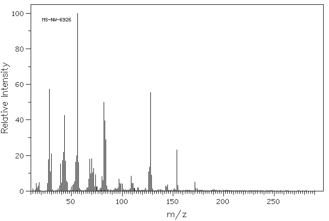
质谱MS
-
碳谱13CNMR
-
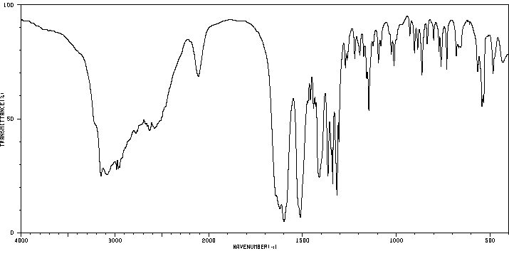
红外IR
-
拉曼Raman
-
峰位数据
-
峰位匹配
-
表征信息
同类化合物
(甲基3-(二甲基氨基)-2-苯基-2H-azirene-2-羧酸乙酯)
(±)-盐酸氯吡格雷
(±)-丙酰肉碱氯化物
(d(CH2)51,Tyr(Me)2,Arg8)-血管加压素
(S)-(+)-α-氨基-4-羧基-2-甲基苯乙酸
(S)-阿拉考特盐酸盐
(S)-赖诺普利-d5钠
(S)-2-氨基-5-氧代己酸,氢溴酸盐
(S)-2-[[[(1R,2R)-2-[[[3,5-双(叔丁基)-2-羟基苯基]亚甲基]氨基]环己基]硫脲基]-N-苄基-N,3,3-三甲基丁酰胺
(S)-2-[3-[(1R,2R)-2-(二丙基氨基)环己基]硫脲基]-N-异丙基-3,3-二甲基丁酰胺
(S)-1-(4-氨基氧基乙酰胺基苄基)乙二胺四乙酸
(S)-1-[N-[3-苯基-1-[(苯基甲氧基)羰基]丙基]-L-丙氨酰基]-L-脯氨酸
(R)-乙基N-甲酰基-N-(1-苯乙基)甘氨酸
(R)-丙酰肉碱-d3氯化物
(R)-4-N-Cbz-哌嗪-2-甲酸甲酯
(R)-3-氨基-2-苄基丙酸盐酸盐
(R)-1-(3-溴-2-甲基-1-氧丙基)-L-脯氨酸
(N-[(苄氧基)羰基]丙氨酰-N〜5〜-(diaminomethylidene)鸟氨酸)
(6-氯-2-吲哚基甲基)乙酰氨基丙二酸二乙酯
(4R)-N-亚硝基噻唑烷-4-羧酸
(3R)-1-噻-4-氮杂螺[4.4]壬烷-3-羧酸
(3-硝基-1H-1,2,4-三唑-1-基)乙酸乙酯
(2S,4R)-Boc-4-环己基-吡咯烷-2-羧酸
(2S,3S,5S)-2-氨基-3-羟基-1,6-二苯己烷-5-N-氨基甲酰基-L-缬氨酸
(2S,3S)-3-((S)-1-((1-(4-氟苯基)-1H-1,2,3-三唑-4-基)-甲基氨基)-1-氧-3-(噻唑-4-基)丙-2-基氨基甲酰基)-环氧乙烷-2-羧酸
(2S)-2,6-二氨基-N-[4-(5-氟-1,3-苯并噻唑-2-基)-2-甲基苯基]己酰胺二盐酸盐
(2S)-2-氨基-N,3,3-三甲基-N-(苯甲基)丁酰胺
(2S)-2-氨基-3-甲基-N-2-吡啶基丁酰胺
(2S)-2-氨基-3,3-二甲基-N-(苯基甲基)丁酰胺,
(2S)-2-氨基-3,3-二甲基-N-2-吡啶基丁酰胺
(2S,4R)-1-((S)-2-氨基-3,3-二甲基丁酰基)-4-羟基-N-(4-(4-甲基噻唑-5-基)苄基)吡咯烷-2-甲酰胺盐酸盐
(2R,3'S)苯那普利叔丁基酯d5
(2R)-2-氨基-3,3-二甲基-N-(苯甲基)丁酰胺
(2-氯丙烯基)草酰氯
(1S,3S,5S)-2-Boc-2-氮杂双环[3.1.0]己烷-3-羧酸
(1R,5R,6R)-5-(1-乙基丙氧基)-7-氧杂双环[4.1.0]庚-3-烯-3-羧酸乙基酯
(1R,4R,5S,6R)-4-氨基-2-氧杂双环[3.1.0]己烷-4,6-二羧酸
齐特巴坦
齐德巴坦钠盐
齐墩果-12-烯-28-酸,2,3-二羟基-,苯基甲基酯,(2a,3a)-
齐墩果-12-烯-28-酸,2,3-二羟基-,羧基甲基酯,(2a,3b)-(9CI)
黄酮-8-乙酸二甲氨基乙基酯
黄荧菌素
黄体生成激素释放激素(1-6)
黄体生成激素释放激素 (1-5) 酰肼
黄体瑞林
麦醇溶蛋白
麦角硫因
麦芽聚糖六乙酸酯
麦根酸

联系我们


关注我们

公众号
