
4-氨基安替比林盐酸盐 | 22198-72-7
中文名称
4-氨基安替比林盐酸盐
中文别名
4-氢氯化氨基安替比林
英文名称
4-aminoantipyrine hydrochloride
英文别名
4-amino-1,5-dimethyl-2-phenylpyrazol-3-one;hydron;chloride

CAS
22198-72-7
化学式
C11H13N3O*ClH
mdl
MFCD00042012
分子量
239.705
InChiKey
UZSCVCWALGRUTR-UHFFFAOYSA-N
BEILSTEIN
——
EINECS
——
-
物化性质
-
计算性质
-
ADMET
-
安全信息
-
SDS
-
制备方法与用途
-
上下游信息
-
文献信息
-
表征谱图
-
同类化合物
-
相关功能分类
-
相关结构分类
物化性质
-
熔点:220-225 °C
-
稳定性/保质期:
在常温常压下稳定,应避免接触强氧化剂、强酸、酸性氯化物以及酸酐。
计算性质
-
辛醇/水分配系数(LogP):0.33
-
重原子数:16
-
可旋转键数:1
-
环数:2.0
-
sp3杂化的碳原子比例:0.181
-
拓扑面积:49.6
-
氢给体数:2
-
氢受体数:3
安全信息
-
危险品标志:Xn
-
危险类别码:R36/37/38
-
海关编码:2933199090
-
安全说明:S26,S36/37/39
-
危险性防范说明:P264,P270,P280,P301+P312+P330,P302+P352+P332+P313+P362+P364,P305+P351+P338+P337+P313,P501
-
危险性描述:H302,H315,H319
-
储存条件:将物品存放在密封的容器中,并储存在阴凉、干燥的地方。
SDS
4-氨基安替比林盐酸盐[用于生化研究] 修改号码:5
模块 1. 化学品
产品名称: 4-AminoaNTipyrine Hydrochloride [for Biochemical Research]
修改号码: 5
模块 2. 危险性概述
GHS分类
物理性危害 未分类
健康危害
急性毒性(经口) 第4级
皮肤腐蚀/刺激 第2级
严重损伤/刺激眼睛 2A类
环境危害 未分类
GHS标签元素
图标或危害标志
信号词 警告
危险描述 吞咽有害。
造成皮肤刺激
造成严重眼刺激
防范说明
[预防] 使用本产品时切勿吃东西,喝水或吸烟。
处理后要彻底清洗双手。
穿戴防护手套/护目镜/防护面具。
[急救措施] 食入:若感不适,呼叫解毒中心/医生。漱口。
眼睛接触:用水小心清洗几分钟。如果方便,易操作,摘除隐形眼镜。继续冲洗。
眼睛接触:求医/就诊
皮肤接触:用大量肥皂和水轻轻洗。
若皮肤刺激:求医/就诊。
脱掉被污染的衣物,清洗后方可重新使用。
[废弃处置] 根据当地政府规定把物品/容器交与工业废弃处理机构。
4-氨基安替比林盐酸盐[用于生化研究] 修改号码:5
模块 3. 成分/组成信息
单一物质/混和物 单一物质
化学名(中文名): 4-氨基安替比林盐酸盐[用于生化研究]
百分比: >98.0%(LC)(T)
CAS编码: 22198-72-7
俗名:
4-AA·HCl
分子式: C11H13N3O·HCl
模块 4. 急救措施
吸入: 将受害者移到新鲜空气处,保持呼吸通畅,休息。若感不适请求医/就诊。
皮肤接触: 立即去除/脱掉所有被污染的衣物。用大量肥皂和水轻轻洗。
若皮肤刺激或发生皮疹:求医/就诊。
眼睛接触: 用水小心清洗几分钟。如果方便,易操作,摘除隐形眼镜。继续清洗。
如果眼睛刺激:求医/就诊。
食入: 若感不适,呼叫解毒中心/医生。漱口。
紧急救助者的防护: 救援者需要穿戴个人防护用品,比如橡胶手套和气密性护目镜。
模块 5. 消防措施
合适的灭火剂: 干粉,泡沫,雾状水,二氧化碳
特殊危险性: 小心,燃烧或高温下可能分解产生毒烟。
特定方法: 从上风处灭火,根据周围环境选择合适的灭火方法。
非相关人员应该撤离至安全地方。
周围一旦着火:如果安全,移去可移动容器。
消防员的特殊防护用具: 灭火时,一定要穿戴个人防护用品。
模块 6. 泄漏应急处理
个人防护措施,防护用具, 使用个人防护用品。远离溢出物/泄露处并处在上风处。
紧急措施: 泄露区应该用安全带等圈起来,控制非相关人员进入。
环保措施: 防止进入下水道。
控制和清洗的方法和材料: 清扫收集粉尘,封入密闭容器。注意切勿分散。附着物或收集物应该立即根据合适的
法律法规处置。
模块 7. 操作处置与储存
处理
技术措施: 在通风良好处进行处理。穿戴合适的防护用具。防止粉尘扩散。处理后彻底清洗双手
和脸。
注意事项: 如果粉尘或浮质产生,使用局部排气。
操作处置注意事项: 避免接触皮肤、眼睛和衣物。
贮存
储存条件: 保持容器密闭。存放于凉爽、阴暗处。
远离不相容的材料比如氧化剂存放。
包装材料: 依据法律。
模块 8. 接触控制和个体防护
工程控制: 尽可能安装封闭体系或局部排风系统,操作人员切勿直接接触。同时安装淋浴器和洗
眼器。
个人防护用品
呼吸系统防护: 防尘面具。依据当地和政府法规。
手部防护: 防护手套。
4-氨基安替比林盐酸盐[用于生化研究] 修改号码:5
模块 8. 接触控制和个体防护
眼睛防护: 安全防护镜。如果情况需要,佩戴面具。
皮肤和身体防护: 防护服。如果情况需要,穿戴防护靴。
模块 9. 理化特性
固体
外形(20°C):
外观: 晶体-粉末
颜色: 微浅黄色-浅红黄色
气味: 无资料
pH: 无数据资料
熔点: 无资料
沸点/沸程 无资料
闪点: 无资料
爆炸特性
爆炸下限: 无资料
爆炸上限: 无资料
密度: 无资料
溶解度:
[水] 无资料
[其他溶剂] 无资料
模块 10. 稳定性和反应性
化学稳定性: 一般情况下稳定。
危险反应的可能性: 未报道特殊反应性。
须避免接触的物质 氧化剂
危险的分解产物: 一氧化碳, 二氧化碳, 氮氧化物 (NOx), 氯化氢
模块 11. 毒理学信息
急性毒性: 无资料
对皮肤腐蚀或刺激: 无资料
对眼睛严重损害或刺激: 无资料
生殖细胞变异原性: 无资料
致癌性:
IARC = 无资料
NTP = 无资料
生殖毒性: 无资料
模块 12. 生态学信息
生态毒性:
鱼类: 无资料
甲壳类: 无资料
藻类: 无资料
残留性 / 降解性: 无资料
潜在生物累积 (BCF): 无资料
土壤中移动性
log水分配系数: 无资料
土壤吸收系数 (Koc): 无资料
亨利定律 无资料
constaNT(PaM3/mol):
4-氨基安替比林盐酸盐[用于生化研究] 修改号码:5
模块 13. 废弃处置
如果可能,回收处理。请咨询当地管理部门。建议在可燃溶剂中溶解混合,在装有后燃和洗涤装置的化学焚烧炉中
焚烧。废弃处置时请遵守国家、地区和当地的所有法规。
模块 14. 运输信息
联合国分类: 与联合国分类标准不一致
UN编号: 未列明
模块 15. 法规信息
《危险化学品安全管理条例》(2002年1月26日国务院发布,2011年2月16日修订): 针对危险化学品的安全使用、
生产、储存、运输、装卸等方面均作了相应的规定。
模块16 - 其他信息
N/A
制备方法与用途
纯化方法
称取2克4-氨基安替比林,用水溶解后定容至100毫升,并将溶液置于冰箱中保存。纯化步骤如下:
向上述浓度为2%(质量分数)的4-氨基安替比林溶液中加入10克漂白土,搅拌均匀后静置。随后进行离心分离,收集滤液,得到纯化的4-氨基安替比林溶液。
根据中华人民共和国国家环境保护标准HJ503-2009《水质挥发酚的测定 4-氨基安替比林分光光度法》中的18.4条方法,分别测定了纯化前后溶液的空白吸光度值,分别为0.161和0.049。
合成方法游离碱合成步骤如下:
- 将安替比林与50%硫酸混合配制成溶液。
- 在亚硝化反应器中同时加入上述溶液以及亚硝酸钠溶液,并控制两者的流量,确保反应温度在45-50℃之间。在搅拌的同时进行反应,通过碘粉淀粉试纸检测反应终点以调节水的流动速率。
- 反应生成的亚硝基安替比林立即流入还原罐,在该罐内与预先配置好的还原剂水溶液发生反应。
- 从反应液中取样测定pH值。当pH值达到5.4-5.8之间时,表示还原结束;接下来将pH值调节至5.8-6.0,并升温至100℃进行3小时的水解处理。之后将温度降至80℃,用液氨中和至pH7-7.5后静置分层。
- 分离去废水后,得到4-氨基安替比林油。将其压入结晶罐,并进行搅拌、冷却结晶和过滤操作,最终得到4-氨基安替比林产品。
反应信息
-
作为反应物:描述:4-氨基安替比林盐酸盐 在 盐酸 、 水 、 sodium hydride 、 N,N-二异丙基乙胺 、 nickel dichloride 作用下, 以 四氢呋喃 、 CH3Cl 、 mineral oil 为溶剂, 生成 4-(甲氨基)安替比林参考文献:名称:NMR-Derived Models of Amidopyrine and Its Metabolites in Complexes with Rabbit Cytochrome P450 2B4 Reveal a Structural Mechanism of Sequential N-Dealkylation摘要:To understand the molecular basis of sequential N-dealkylation by cytochrome P450 2B enzymes, we studied the binding of amidopyrine (AP) as well as the metabolites of this reaction, desmethylamidopyrine (DMAP) and aminoantipyrine (AAP), using the X-ray crystal structure of rabbit P450 2B4 and two nuclear magnetic resonance (NMR) techniques: saturation transfer difference (STD) spectroscopy and longitudinal (T-1) relaxation NMR. Results of STD NMR of AP and its metabolites bound to P450 2B4 were similar, suggesting that they occupy similar niches within the enzyme's active site. The model-dependent relaxation rates (R-M) determined from T-1 relaxation NMR of AP and DMAP suggest that the N-linked methyl is closest to the heme. To determine the orientation(s) of AP and its metabolites within the P450 2B4 active site, we used distances calculated from the relaxation rates to constrain the metabolites to the X-ray crystal structure of P450 2B4. Simulated annealing of the complex revealed that the metabolites do indeed occupy similar hydrophobic pockets within the active site, while the N-linked methyls are free to rotate between two binding modes. From these bound structures, a model of N-demethylation in which the N-linked methyl functional groups rotate between catalytic and noncatalytic positions was developed. This study is the first to provide a structural model of a drug and its metabolites complexed to a c-ytochrome P450 based on NMR and to provide a structural mechanism for how a drug can undergo sequential oxidations without unbinding. The rotation of the amide functional group might represent a common structural mechanism for N-dealkylation reactions for other drugs such as the local anesthetic lidocaine.DOI:10.1021/bi101797v
-
作为产物:描述:参考文献:名称:萘酚-2-醇偶氮衍生物的合成、晶体结构、溶剂致变色特性和DNA切割活性摘要:摘要 4-氨基安替比林的萘酚偶氮化合物C21H18N4O2 在室温下通过DMF 缓慢蒸发生长了晶体。生长的晶体通过 FT-IR、UV-Vis、13C 和 1H NMR 光谱表征。通过单晶X射线衍射数据分析分子结构。化合物4-[(E)-(3-羟基萘-2-基)二氮烯基]-1,5-二甲基-2-苯基-1,2-二氢-3H-吡唑-3-one在单斜晶系P21中结晶/c 空间群具有单元尺寸 a = 18.7902 (12) A, b = 6.4128 (5) A, c = 14.7923 (11) A 和 β = 91.229 (2)°。染料的溶剂致变色是根据九种溶剂中吸收最大值的波长来评估的。由于分子内氢键的存在,腙形式在大多数极性质子和极性非质子溶剂中占主导地位。还报告了 pH 值对染料可见吸收光谱的影响。还强调了光谱值与溶剂参数的拟合以及与 Kamlet-Abboud-Taft 方程的相关性。标题化合物与DOI:10.1016/j.molstruc.2018.11.093
文献信息
-
Immunoassay element申请人:KONICA CORPORATION公开号:EP0327918A1公开(公告)日:1989-08-16An immunoassay element for assaying a target substance in a fluid sample by utilizing homogeneous competitive reaction of the target substance and a labelled substance which is a target substance or an analogue thereof labelled with a marker, with a substance which specifically reacts with both the target substance and the labelled substance. The immunoassay element comprises, in the order mentioned from the top to the bottom, the layers of a) a fibrous porous spreading layer; b) a non-fibrous porous reaction layer comprising the substance which specifically reacts with both the target substance and the labelled substance; and c) a non-porous layer comprising a macromolecular substance in which the labelled substance is incorporated. The labelled substance gives a signal which is changed when the labelled substance is bound to the substance which specifically reacts with both the target substance and the labelled substance.一种免疫测定元件,用于利用目标物质和标记物质的同质竞争反应来测定流体样品中的目标物质,标记物质是用标记物标记的目标物质或其类似物,与目标物质和标记物质均发生特异性反应的物质。免疫测定元件按从上到下的顺序包括以下各层:a) 纤维状多孔铺展层;b) 非纤维状多孔反应层,包括与目标物质和标记物质发生特异反应的物质;c) 非多孔层,包括大分子物质,其中含有标记物质。当被标记的物质与能与目标物质和标记物质发生特异性反应的物质结合时,标记物质发出的信号会发生变化。
-
Analyte concentration detection devices and methods申请人:Intuity Medical, Inc.公开号:EP2322914A1公开(公告)日:2011-05-18Arrangements for the detection of the presence and/or concentration of an analyte in a sample of bodily fluid include diffuse transmission, diffuse reflection and edge or waveguide illumination arrangements. A vertical flow assay and/or detection component (100) is provided in the form of an array of optical detection elements (24).用于检测体液样本中是否存在分析物和/或分析物浓度的装置包括漫反射透射、漫反射反射和边缘或波导照明装置。垂直流分析和/或检测组件(100)以光学检测元件阵列(24)的形式提供。
-
NUCLEIC ACID ELEMENT FOR USE IN ANALYSIS, AND ANALYTICAL METHOD, ANALYTICAL REAGENT AND ANALYTICAL INSTRUMENT USING SAME申请人:NEC Soft, Ltd.公开号:EP2463660A1公开(公告)日:2012-06-13The technique by which simple analysis of an intended subject to be analyzed can be carried out is provided, In this technique, a nucleic acid element 16 for use in analysis including: a first nucleic acid part 12; and a second nucleic acid part 13 is used. In the nucleic acid element 16, the first nucleic acid part 12 is a binding part that can bind to a subject 11 to be analyzed, and the second nucleic acid part 13 is a labeling part that can distinguish between binding and non-binding of the first nucleic acid part 12 to the subject 11. It is preferred that the first nucleic acid part 12 is an aptamer against the subject 11. The subject 11 can be analyzed easily by using the nucleic acid element 16, binding the subject 11 to the first nucleic acid part 12, and then analyzing the binding with the second nucleic acid part 13.在该技术中,使用了一种用于分析的核酸元件 16,包括:第一核酸部分 12 和第二核酸部分 13。在核酸元件 16 中,第一核酸部分 12 是可与待分析对象 11 结合的结合部分,第二核酸部分 13 是可区分第一核酸部分 12 与对象 11 结合与否的标记部分。第一核酸部分 12 最好是针对受试者 11 的适配体。使用核酸元件 16,将受试者 11 与第一核酸部分 12 结合,然后用第二核酸部分 13 分析结合情况,就可以很容易地分析受试者 11。
-
DETECTION TOOL, AND DETECTION SYSTEM申请人:NEC Soft, Ltd.公开号:EP2657330A1公开(公告)日:2013-10-30The present invention provides a detection instrument capable of easily detecting an intended detection object without any skilled technique. The detection instrument (1) of the present invention includes a detection portion (12), a detection reagent which develops a color by specifically reacting with a detection object in a sample is placed in the detection portion (12), positional information of the detection reagent in the detection portion (12) is information on the detection object, and color development of the detection reagent can be optically read. It is preferred that a bar code is formed in the detection portion (12), and the detection reagent is placed as a part of the bar code.本发明提供了一种检测仪器,无需任何熟练技术即可轻松检测到预定的检测对象。本发明的检测仪器(1)包括一个检测部分(12),检测部分(12)中放置了通过与样品中的检测对象发生特异性反应而显色的检测试剂,检测部分(12)中检测试剂的位置信息是检测对象的信息,检测试剂的显色情况可以通过光学读取。最好在检测部分(12)中形成条形码,并将检测试剂作为条形码的一部分放置。
-
ANALYTICAL DEVICE AND ANALYTICAL METHOD申请人:NEC Soft, Ltd.公开号:EP2657702A1公开(公告)日:2013-10-30The present invention provides a technique capable of simply analyzing a target to be analyzed. An analytical device of the present invention includes a basal plate; a nucleic acid element; and a detection section of detecting a signal. The nucleic acid element and the detection section are arranged on the basal plate. The nucleic acid element includes a first nucleic acid molecule and a second nucleic acid molecule. The first nucleic acid molecule is a nucleic acid molecule capable of binding to a target. The second nucleic acid molecule is a nucleic acid molecule capable of binding to streptavidin. When the target does not bind to the first nucleic acid molecule, a binding capacity of the second nucleic acid molecule to the streptavidin is inactivated. When the target binds to the first nucleic acid molecule, a binding capacity of the second nucleic acid molecule to the streptavidin is activated. The detection section detects binding between the second nucleic acid molecule and the streptavidin. The target is bound to the first nucleic acid molecule, so that the streptavidin is bound to the second nucleic acid molecule. Thus, the target can be analyzed through detecting the binding between the second nucleic acid molecule and the streptavidin using the detection device.
表征谱图
-
氢谱1HNMR
-
质谱MS
-
碳谱13CNMR
-
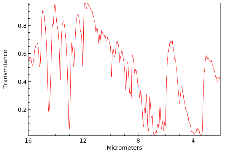
红外IR
-
拉曼Raman
-
峰位数据
-
峰位匹配
-
表征信息
同类化合物
(SP-4-1)-二氯双(1-苯基-1H-咪唑-κN3)-钯
(5aS,6R,9S,9aR)-5a,6,7,8,9,9a-六氢-6,11,11-三甲基-2-(2,3,4,5,6-五氟苯基)-6,9-甲基-4H-[1,2,4]三唑[3,4-c][1,4]苯并恶嗪四氟硼酸酯
(5-氨基-1,3,4-噻二唑-2-基)甲醇
齐墩果-2,12-二烯[2,3-d]异恶唑-28-酸
黄曲霉毒素H1
高效液相卡套柱
非昔硝唑
非布索坦杂质Z19
非布索坦杂质T
非布索坦杂质K
非布索坦杂质E
非布索坦杂质D
非布索坦杂质67
非布索坦杂质65
非布索坦杂质64
非布索坦杂质61
非布索坦代谢物67M-4
非布索坦代谢物67M-2
非布索坦代谢物 67M-1
非布索坦-D9
非布索坦
非唑拉明
雷非那酮-d7
雷西那德杂质2
雷西纳德杂质L
雷西纳德杂质H
雷西纳德杂质B
雷西纳德
雷西奈德杂质
阿西司特
阿莫奈韦
阿考替胺杂质9
阿米苯唑
阿米特罗13C2,15N2
阿瑞匹坦杂质
阿格列扎
阿扎司特
阿尔吡登
阿塔鲁伦中间体
阿培利司N-1
阿哌沙班杂质26
阿哌沙班杂质15
阿可替尼
阿作莫兰
阿佐塞米
镁(2+)(Z)-4'-羟基-3'-甲氧基肉桂酸酯
锌1,2-二甲基咪唑二氯化物
锌(II)(苯甲醇)(四苯基卟啉)
锌(II)(正丁醇)(四苯基卟啉)
锌(II)(异丁醇)(四苯基卟啉)

联系我们


关注我们

公众号

