Ammonia solutions (containing more than 35% but not more than 50% ammonia) appears as a clear colorless liquid consisting of ammonia dissolved in water. Corrosive to tissue and metals. Although ammonia is lighter than air, the vapors from a leak will initially hug the ground. Long term exposure to low concentrations or short term exposure to high concentrations may result in adverse health conditions from inhalation. Prolonged exposure of containers to fire or heat may result in their violent rupturing and rocketing.
颜色/状态:
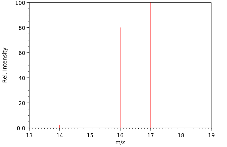
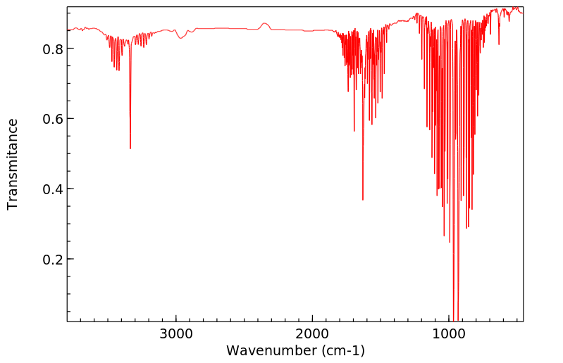
Colorless gas
气味:
Sharp, cloying, repellent
蒸汽密度:
0.6 (EPA, 1998) (Relative to Air)
蒸汽压力:
Vapor pressure: 1 Pa at -139 °C, 10 Pa at -127 °C, 100 Pa at -112 °C; 1 kPa at -94.5 °C (solids); 10 kPa at -71.3 °C, 100 kPa at -33.6 °C (liquid)
亨利常数:
Henry's Law constant = 1.61X10⁻⁵ atm cu-m/mole at 25 °C
Healthy hepatocytes detoxify ammonia where hepatic glutaminase, glutamine synthetase and the urea cycle enzymes act as major enzymes for ammonia metabolism. Ammonia is converted to urea in the liver and other tissues. Glutaminase and glutamine synthetase catalyze the condensation of ammonia with glutamate to glutamine, which is a common nontoxic carrier of ammonia. In case of hepatic dysfunction or impairment, detoxification capacity decreases and may cause severe pathologies from hyperammonemia, such as hepatic encephalopathy.
Human adults produce around 1000 mmol of ammonia daily. Some is reutilized in biosynthesis. The remainder is waste and neurotoxic. Eventually most is excreted in urine as urea, together with ammonia used as a buffer. In extrahepatic tissues, ammonia is incorporated into nontoxic glutamine and released into blood. Large amounts are metabolized by the kidneys and small intestine. In the intestine, this yields ammonia, which is sequestered in portal blood and transported to the liver for ureagenesis, and citrulline, which is converted to arginine by the kidneys. The amazing developments in NMR imaging and spectroscopy and molecular biology have confirmed concepts derived from early studies in animals and cell cultures. The processes involved are exquisitely tuned. When they are faulty, ammonia accumulates. Severe acute hyperammonemia causes a rapidly progressive, often fatal, encephalopathy with brain edema. Chronic milder hyperammonemia causes a neuropsychiatric illness. Survivors of severe neonatal hyperammonemia have structural brain damage. Proposed explanations for brain edema are an increase in astrocyte osmolality, generally attributed to glutamine accumulation, and cytotoxic oxidative/nitrosative damage. However, ammonia neurotoxicity is multifactorial, with disturbances also in neurotransmitters, energy production, anaplerosis, cerebral blood flow, potassium, and sodium. Around 90% of hyperammonemic patients have liver disease. Inherited defects are rare. They are being recognized increasingly in adults. Deficiencies of urea cycle enzymes, citrin, and pyruvate carboxylase demonstrate the roles of isolated pathways in ammonia metabolism. Phenylbutyrate is used routinely to treat inherited urea cycle disorders, and its use for hepatic encephalopathy is under investigation. /Hyperammonemia/
The inhibitory effects of ammonia on two different degradation pathways of methanogenic acetate were evaluated using a pure culture (Methanosaeta thermophila strain PT) and defined co-culture (Methanothermobacter thermautotrophicus strain TM and Thermacetogenium phaeum strain PB), which represented aceticlastic and syntrophic methanogenesis, respectively. Growth experiments with high concentrations of ammonia clearly demonstrated that sensitivity to ammonia stress was markedly higher in M. thermophila PT than in the syntrophic co-culture. M. thermophila PT also exhibited higher sensitivity to high pH stress, which indicated that an inability to maintain pH homeostasis is an underlying cause of ammonia inhibition. Methanogenesis was inhibited in the resting cells of M. thermophila PT with moderate concentrations of ammonia, suggesting that the inhibition of enzymes involved in methanogenesis may be one of the major factors responsible for ammonia toxicity. Transcriptomic analysis revealed a broad range of disturbances in M. thermophila PT cells under ammonia stress conditions, including protein denaturation, oxidative stress, and intracellular cation imbalances. The results of the present study clearly demonstrated that syntrophic acetate degradation dominated over aceticlastic methanogenesis under ammonia stress conditions, which is consistent with the findings of previous studies on complex microbial community systems. Our results also imply that the co-existence of multiple metabolic pathways and their different sensitivities to stress factors confer resiliency on methanogenic processes.
Recently, spatial-temporal/metabolic mathematical models have been established that allow the simulation of metabolic processes in tissues. We applied these models to decipher ammonia detoxification mechanisms in the liver. An integrated metabolic-spatial-temporal model was used to generate hypotheses of ammonia metabolism. Predicted mechanisms were validated using time-resolved analyses of nitrogen metabolism, activity analyses, immunostaining and gene expression after induction of liver damage in mice. Moreover, blood from the portal vein, liver vein and mixed venous blood was analyzed in a time dependent manner. Modeling revealed an underestimation of ammonia consumption after liver damage when only the currently established mechanisms of ammonia detoxification were simulated. By iterative cycles of modeling and experiments, the reductive amidation of alpha-ketoglutarate (alpha-KG) via glutamate dehydrogenase (GDH) was identified as the lacking component. GDH is released from damaged hepatocytes into the blood where it consumes ammonia to generate glutamate, thereby providing systemic protection against hyperammonemia. This mechanism was exploited therapeutically in a mouse model of hyperammonemia by injecting GDH together with optimized doses of cofactors. Intravenous injection of GDH (720 U/kg), alpha-KG (280 mg/kg) and NADPH (180 mg/kg) reduced the elevated blood ammonia concentrations (>200 uM) to levels close to normal within only 15 min. If successfully translated to patients the GDH-based therapy might provide a less aggressive therapeutic alternative for patients with severe hyperammonemia. /Hyperammonemia/
The rodent liver eliminates toxic ammonia. In mammals, three enzymes (or enzyme systems) are involved in this process: glutaminase, glutamine synthetase and the urea cycle enzymes, represented by carbamoyl phosphate synthetase. The distribution of these enzymes for optimal ammonia detoxification was determined by numerical optimization. This in silico approach predicted that the enzymes have to be zonated in order to achieve maximal removal of toxic ammonia and minimal changes in glutamine concentration. Using 13 compartments, representing hepatocytes, the following predictions were generated: glutamine synthetase is active only within a narrow pericentral zone. Glutaminase and carbamoyl phosphate synthetase are located in the periportal zone in a non-homogeneous distribution. This correlates well with the paradoxical observation that in a first step glutamine-bound ammonia is released (by glutaminase) although one of the functions of the liver is detoxification by ammonia fixation. The in silico approach correctly predicted the in vivo enzyme distributions also for non-physiological conditions (e.g. starvation) and during regeneration after tetrachloromethane (CCl4) intoxication. Metabolite concentrations of glutamine, ammonia and urea in each compartment, representing individual hepatocytes, were predicted. Finally, a sensitivity analysis showed a striking robustness of the results. These bioinformatics predictions were validated experimentally by immunohistochemistry and are supported by the literature. In summary, optimization approaches like the one applied can provide valuable explanations and high-quality predictions for in vivo enzyme and metabolite distributions in tissues and can reveal unknown metabolic functions.
IDENTIFICATION AND USE: Ammonia is a colorless gas or liquid. Ammonia is used in the production of ammonium sulfate and ammonium nitrate for fertilizers; and in the manufacture of nitric acid, soda, synthetic urea, synthetic fibers, dyes, and plastics. Ammonia, or dissociated ammonia, is used in such metal treating operations as nitriding, carbo-nitriding, bright annealing, furnace brazing, sintering, sodium hydride descaling, atomic hydrogen welding, and other applications where protective atmospheres are required. The petroleum industry utilizes anhydrous ammonia in neutralizing the acid constituents of crude oil and in protecting equipment such as bubble plate towers, heat exchangers, condensers, and storage tanks from corrosion. It is also used as medication. Ammonia in an aqueous environment exists in equilibrium between ionized ammonium cation and the non-ionized ammonia. This equilibrium can be affected by buffers, pH, temperature, and salinity. Thus, in many cases it is not possible to assign the associated toxicity to the ionized or non-ionized form of the ammonia-nitrogen. HUMAN EXPOSURE AND TOXICITY: Studies using low levels of ammonia show that inhaled ammonia is temporarily dissolved in the mucus of the upper respiratory tract, and then a high percentage of it is released back into the expired air. Following exposure to 500 ppm ammonia for 10-27 min, healthy male subjects eliminated 70-80% of the inspired ammonia by this route. Short term exposure: eye or skin contact with ammonia can cause irritation, burns, frostbite (anhydrous), and permanent damage. Irritates the respiratory tract causing coughing, wheezing, and shortness of breath. Higher exposure can cause pulmonary edema, a medical emergency, that can be delayed for several hours and is life-threatening. Exposure can cause headache, loss of sense of smell, nausea, and vomiting. Inhalation: nose and throat irritation have been reported at 72 ppm after 5 min exposure. Exposures of 500 ppm for 30 min have caused upper respiratory irritation, tearing, increased pulse rate, and blood pressure. Death has been reported after an exposure to 10,000 ppm for an unknown duration. Skin: Solutions of 2% ammonia can cause burns and blisters after 15 min of exposure. These burns may be slow to heal. Anhydrous ammonia may cause skin to freeze. Eyes: Levels of 70 ppm (gas) have caused eye irritation. If not flushed with water immediately, contact with eye may cause partial or complete blindness. Ingestion: ammonia will cause pain if swallowed and burning of the throat and stomach. May cause vomiting. One teaspoon of 28% aqua ammonia may cause death. Long term exposure: repeated exposure can cause chronic eye, nose, and throat irritation. Repeated lung irritation can result in bronchitis with coughing, shortness of breath, and phlegm. Analysis of blood samples from 22 workers exposed to ammonia in a fertilizer factory and 42 control workers not exposed to ammonia showed increased frequency of chromosomal aberrations (CAs) and sister chromatid exchanges (SCEs), increased mitotic index (MI), and increased frequency of CAs and SCEs with increasing length of exposure. ANIMAL STUDIES: Analysis of endogenous ammonia levels in the expired air of rats showed concentrations ranging from 10-353 ppb (mean = 78 ppb) in nose-breathing animals. The quantitative difference between inspired and expired ammonia suggests that small amounts are absorbed across the nasopharyngeal membranes into the systemic circulation. Absorbed ammonia is excreted by the kidneys as urea and urinary ammonium compounds, as urea in feces, and as components of sweat. Toxic levels do not develop as a result of chronic inhalation exposure because the body has multiple effective mechanisms for detoxifying and excreting it. Cardiovascular changes that may be analogous to those observed in humans have been observed in rabbits exposed to high concentrations of ammonia. Bradycardia was seen at 2,500 ppm, and hypertension and cardiac arrhythmias leading to cardiovascular collapse followed acute exposures to concentrations exceeding 5,000 ppm. Pathological correlates for these effects have not been demonstrated. Atrophy of pericardial fat has been observed in mice exposed to 4,000 ppm ammonia. Hepatic effects are usually not seen in animals exposed to ammonia gas. Liver necrosis has been observed following acute lethal exposure of mice to 3,440 ppm ammonia for 1 hour. Levels of 170 ppm of ammonia vapor caused mild changes in the spleens, kidneys, and livers of guinea pigs. Static exposures of cats and rabbits for 1 hr at 7000 mg/cu m resulted in the death of approx 50%. Postmortem exam showed severe effects on the upper respiratory tract. Less severe effects in the lower respiratory tract included damage to bronchioles and alveolar congestion, edema, atelectasis, hemorrhage, emphysema, and fluid. The search for the peripheral toxins responsible for the CNS impairment present in hepatic encephalopathy has shown that the administration of ammonia in normal rats reproduced behavioral and electrophysiological changes similar to those seen in galactosamine induced encephalopathy. No statistically significant differences were noted in ovarian or uterine weights of pigs exposed to about 7 or 35 ppm ammonia for 6 weeks. Female pigs that were continuously exposed to about 35 ppm ammonia from 6 weeks before breeding until day 30 of gestation had no statistically significant differences in age at puberty, number of live fetuses, or fetus-to-corpus luteum ratio compared to pigs exposed to only about 7 ppm. No unexposed controls were included in that study. No statistically significant difference in fetal length was evident at 30 days of gestation in offspring of pig dams that were continuously exposed to about 7 or 35 ppm ammonia from 6 weeks before breeding until day 30 of gestation. The mutagenicity of anhydrous ammonia was investigated in a Ames test in S. typhimurium TA98, TA100, TA1535, TA1537 and TA1538, and in E. coli WP2uvrA. The test method was modified appropriately to investigate a volatile test substance. Studies were performed in duplicate in the presence and absence of an exogenous metabolic activation system. No evidence of mutagenicity was seen under the conditions of this assay. ECOTOXICITY STUDIES: Ammonia is an environmental pollutant that is toxic to all aquatic animals. The major sources for atmospheric NH3 are agricultural activities and animal feedlot operations, followed by biomass burning (including forest fires) and to a lesser extent fossil fuel combustion. Close to its sources, acute exposures to NH3 can result in visible foliar injury on vegetation.
The topical damage caused by ammonia is probably due mainly to its alkaline properties. Its high water solubility allows it to dissolve in moisture on the mucous membranes, skin, and eyes, forming ammonium hydroxide. Ammonium hydroxide causes saponification of cell membrane lipids, resulting in cell disruption and death. Additionally, it extracts water from the cells and initiates an inflammatory response, which further damages the surrounding tissues. Excess circulating levels of ammonia (hyperammonemia) can cause serious neurological effects. This is thought to involve the alteration of glutamate metabolism in the brain and resultant increased activation of NMDA receptors, which causes decreased protein kinase C-mediated phosphorylation of Na+/K+ ATPase, increased activity of Na+/K+ ATPase, and depletion of ATP. Ammonia can chemically interact with an internal thiolester bond of
complement 3 (C3). This causes a conformation change in C3, which activates the alternative complement pathway, causing the release of chemoattractants and the assembly of the membrane attack complex of complement. The altered C3 can also bind directly to phagocyte complement receptors, which causes the release of toxic oxygen species. (L958)
来源:Toxin and Toxin Target Database (T3DB)
毒理性
致癌物分类
对人类无致癌性(未列入国际癌症研究机构IARC清单)。
No indication of carcinogenicity to humans (not listed by IARC).
Acute exposure to high levels of ammonia in air may be irritating to skin, eyes, throat, and lungs and cause coughing and burns. Lung damage and death may occur after exposure to very high concentrations of ammonia. Swallowing concentrated solutions of ammonia can cause burns in mouth, throat, and stomach. Splashing ammonia into eyes can cause burns and even blindness. (L958)
Chronically high levels of ammonia in the blood are associated with nearly 20 different inborn errors of metabolism including: 3-Hydroxy-3-Methylglutaryl-CoA Lyase Deficiency, Argininemia, Argininosuccinic Aciduria, Beta-Ketothiolase Deficiency, Biotinidase deficiency, Carbamoyl Phosphate Synthetase Deficiency, Carnitine-acylcarnitine translocase deficiency, Citrullinemia Type I, Hyperinsulinism-Hyperammonemia Syndrome, Hyperornithinemia-hyperammonemia-homocitrullinuria syndrome, Isovaleric Aciduria, Lysinuric Protein Intolerance, Malonic Aciduria, Methylmalonic Aciduria, Methylmalonic Aciduria Due to Cobalamin-Related Disorders, Propionic acidemia, Pyruvate carboxylase deficiency and Short Chain Acyl CoA Dehydrogenase Deficiency (SCAD Deficiency). Hyperammonemia is one of the metabolic derangements that contribute to hepatic encephalopathy.
来源:Toxin and Toxin Target Database (T3DB)
毒理性
暴露途径
这种物质可以通过吸入被身体吸收。
The substance can be absorbed into the body by inhalation.
来源:ILO-WHO International Chemical Safety Cards (ICSCs)
Ammonia can be absorbed via oral or inhalation route. Inhales ammonia is temporarily dissolved in the mucus of the upper respiratory tract, however the majority of the gas is released back into the air via expiration. In healthy male subjects under exposure to 500 ppm ammonia for 10-27 minutes, about 70-80% of total inspired ammonia was expired. In extrahepatic tissues such as the intestine, ammonia is incorporated into nontoxic glutamine and released into blood, where it is transported to the liver for ureagenesis.
来源:DrugBank
吸收、分配和排泄
消除途径
它主要通过呼出的空气或肾脏排泄来排出体外。
It is mainly excreted through expired air or renal elimination.
Studies suggest that ammonia can be absorbed by the inhalation and oral routes of exposure, but there is less certainty regarding absorption through the skin. Absorption through the eye has been documented. Most of the inhaled ammonia is retained in the upper respiratory tract and is subsequently eliminated in expired air. Almost all of the ammonia produced endogenously in the intestinal tract is absorbed. Exogenous ammonia is also readily absorbed in the intestinal tract. Ammonia that reaches the circulation is widely distributed to all body compartments although substantial first pass metabolism occurs in the liver where it is transformed into urea and glutamine. Ammonia or ammonium ion reaching the tissues is taken up by glutamic acid, which participates in transamination and other reactions. The principal means of excretion of ammonia that reaches the circulation in mammals is as urinary urea; minimal amounts are excreted in the feces and in expired air.
Experiments with volunteers show that ammonia, regardless of its tested concentration in air (range, 57-500 ppm), is almost completely retained in the nasal mucosa (83-92%) during short-term exposure, i.e., up to 120 sec. However, longer-term exposure (10-27 min) to a concentration of 500 ppm resulted in lower retention (4-30%), with 350-400 ppm eliminated in expired air by the end of the exposure period, suggesting an adaptive capability or saturation of the absorptive process. Nasal and pharyngeal irritation, but not tracheal irritation, suggests that ammonia is retained in the upper respiratory tract. Unchanged levels of blood-urea-nitrogen (BUN), non-protein nitrogen, urinary-urea, and urinary-ammonia are evidence of low absorption into the blood. Exposure to common occupational limits of ammonia in air (25 ppm) with 30% retention (and assuming this quantity is absorbed into the blood stream) would yield an increase in blood ammonium concentration of 0.09 mg/L. This calculated rise is only 10% above fasting levels.
Animal data provide supporting evidence for high-percentage nasal retention, thus protecting the lower respiratory tract from exposure (rabbit, dog). Continuous exposure of rats for 24 hr to concentrations up to 32 ppm resulted in significant increase in blood ammonia levels. Exposures to 310-1,157 ppm led to significantly increased blood concentrations of ammonia within 8 hr of exposure initiation, but blood ammonia returned to pre-exposure values within 12 hr of continuous exposure and remained so over the remaining of the 24 hr exposure period. This suggests an adaptive response mechanism may be activated with longerterm exposure.
Catalytic Proton Coupled Electron Transfer from Metal Hydrides to Titanocene Amides, Hydrazides and Imides: Determination of Thermodynamic Parameters Relevant to Nitrogen Fixation
作者:Iraklis Pappas、Paul J. Chirik
DOI:10.1021/jacs.6b08009
日期:2016.10.12
bis(cyclopentadienyl) titanium amides, hydrazides and imides by proton coupled electron transfer (PCET) is described. Twelve different N-H bond dissociation free energies (BDFEs) among the various nitrogen-containing ligands were measured or calculated, and effects of metal oxidation state and N-ligand substituent were determined. Two metal hydride complexes, (η5-C5Me5)(py-Ph)Rh-H (py-Ph = 2-pyridylphenyl
[EN] PYRROLOTRIAZINONE DERIVATIVES AS PI3K INHIBITORS<br/>[FR] DÉRIVÉS DE PYRROLOTRIAZINONE EN TANT QU'INHIBITEURS DES PI3K
申请人:ALMIRALL SA
公开号:WO2014060432A1
公开(公告)日:2014-04-24
New pyrrolotriazinone derivatives having the chemical structure of formula (I), are disclosed; as well as process for their preparation, pharmaceutical compositions comprising them and their use in therapy as inhibitors of Phosphoinositide 3-Kinases (PI3Ks)
Nucleophilic Reactivities of Hydrazines and Amines: The Futile Search for the α-Effect in Hydrazine Reactivities
作者:Tobias A. Nigst、Anna Antipova、Herbert Mayr
DOI:10.1021/jo301497g
日期:2012.9.21
groups increase the reactivities of the α-position of hydrazines, they decrease the reactivities of the β-position. Despite the 102 times lower reactivities of amines and hydrazines in water than in acetonitrile, the relative reactivities of differently substituted amines and hydrazines are almost identical in the two solvents. In both solvents hydrazine has a reactivity similar to that of methylamine.
胺,肼,酰肼和羟胺与苯甲酸铵离子和醌甲基化物的反应动力学通过紫外可见光谱法,在乙腈和水中,使用常规光谱仪,定流和激光闪光技术进行了研究。从这些反应的二阶速率常数k 2,根据线性自由能关系log k 2=s N(N + E)确定亲核参数N和s N。尽管甲基增加了肼的α-位置的反应性,但它们却降低了β-位置的反应性。尽管10 2胺和肼在水中的反应性比乙腈低两倍,不同取代的胺和肼的相对反应性在两种溶剂中几乎相同。在两种溶剂中,肼的反应性都与甲胺类似。这一观察结果暗示,如果考虑到肼具有两个反应中心,Me取代氨中的一个氢比引入氨基更能增加亲核性。log k 2与相应的平衡常数(log K)或布朗斯台德碱度(p K aH)的关系图未显示相对于烷基胺而言,肼或羟胺的亲核性(α效应)增强。
Oxidation of Glycylglycine by KBrO3 in Aqueous Acetic Acid Medium and Comparison with Monomer Glycine: A Kinetic and Mechanistic Study
The kinetics of oxidation reactions of dipeptide glycylglycine (GG) by KBrO3 in aqueous acetic acid medium, under the condition [KBrO3] 3] and fractional order dependence in [GG]. The effect of ionic strength and [AcOH] on rate was studied and thermodynamic parameters were also calculated. Michealis-Menten type mechanism was proposed.
The syntheses, spectral characterization, and crystal structures of 12-(N-methylnitrilium)monocarba-closo-dodecaborate and 12-(N-methylamidinium)-monocarba-closo-dodecaborate ylides are reported. The carborate anion behaves as an inert and non-conjugating negative charge.